


Crystal structure of mouse glyoxalase I complexed with a small molecule inhibitor
Jiang, L.L., Zhou, L.To be published.
Experimental Data Snapshot
Starting Model: experimental
View more details
Entity ID: 1 | |||||
|---|---|---|---|---|---|
| Molecule | Chains | Sequence Length | Organism | Details | Image |
| Lactoylglutathione lyase | 179 | Mus musculus | Mutation(s): 0 Gene Names: Glo1 EC: 4.4.1.5 | 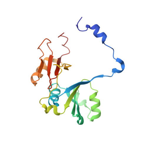 | |
UniProt | |||||
Entity Groups | |||||
| Sequence Clusters | 30% Identity50% Identity70% Identity90% Identity95% Identity100% Identity | ||||
| UniProt Group | Q9CPU0 | ||||
Sequence AnnotationsExpand | |||||
Reference Sequence | |||||
| Ligands 2 Unique | |||||
|---|---|---|---|---|---|
| ID | Chains | Name / Formula / InChI Key | 2D Diagram | 3D Interactions | |
| E1L (Subject of Investigation/LOI) Download:Ideal Coordinates CCD File | E [auth B] | N-[4-(trifluoromethyloxy)phenyl]-1,3,4,9-tetrahydropyrido[3,4-b]indole-2-carbothioamide C19 H16 F3 N3 O S CIBLQSZGHJTWDN-UHFFFAOYSA-N |  | ||
| ZN Download:Ideal Coordinates CCD File | C [auth A], D [auth A] | ZINC ION Zn PTFCDOFLOPIGGS-UHFFFAOYSA-N |  | ||
| Length ( Å ) | Angle ( ˚ ) |
|---|---|
| a = 41.785 | α = 90 |
| b = 64.635 | β = 101.58 |
| c = 65.794 | γ = 90 |
| Software Name | Purpose |
|---|---|
| PHENIX | refinement |
| HKL-3000 | data scaling |
| PDB_EXTRACT | data extraction |
| Coot | model building |


