6KZ8

Crystal structure of plant Phospholipase D alpha complex with phosphatidic acid


Experimental Data Snapshot

  • Method: X-RAY DIFFRACTION
  • Resolution: 2.29 Å
  • R-Value Free: 0.245 
  • R-Value Work: 0.198 
  • R-Value Observed: 0.199 

wwPDB Validation   3D Report Full Report


Ligand Structure Quality Assessment 


This is version 1.3 of the entry. See complete history


Literature

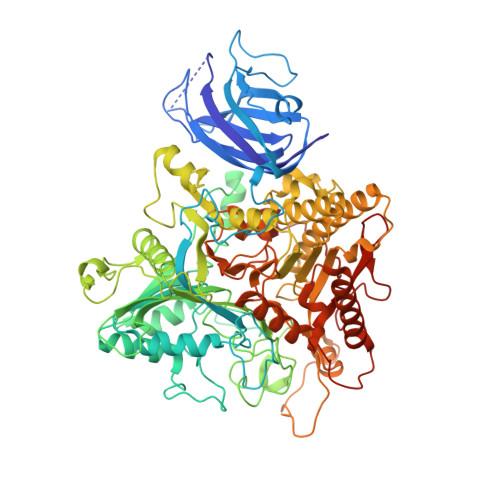
Crystal structure of plant PLD alpha 1 reveals catalytic and regulatory mechanisms of eukaryotic phospholipase D.

Li, J.Yu, F.Guo, H.Xiong, R.Zhang, W.He, F.Zhang, M.Zhang, P.

(2020) Cell Res 30: 61-69

  • DOI: https://doi.org/10.1038/s41422-019-0244-6
  • Primary Citation of Related Structures:  
    6KZ8, 6KZ9

  • PubMed Abstract: 

    Phospholipase D (PLD) hydrolyzes the phosphodiester bond of glycerophospholipids and produces phosphatidic acid (PA), which acts as a second messenger in many living organisms. A large number of PLDs have been identified in eukaryotes, and are viewed as promising targets for drug design because these enzymes are known to be tightly regulated and to function in the pathophysiology of many human diseases. However, the underlying molecular mechanisms of catalysis and regulation of eukaryotic PLD remain elusive. Here, we determined the crystal structure of full-length plant PLDα1 in the apo state and in complex with PA. The structure shows that the N-terminal C2 domain hydrophobically interacts with the C-terminal catalytic domain that features two HKD motifs. Our analysis reveals the catalytic site, substrate-binding mechanism, and a new Ca 2+ -binding site that is required for the activation of PLD. In addition, we tested several efficient small-molecule inhibitors against PLDα1, and suggested a possible competitive inhibition mechanism according to structure-based docking analysis. This study explains many long-standing questions about PLDs and provides structural insights into PLD-targeted inhibitor/drug design.


  • Organizational Affiliation

    National Key Laboratory of Plant Molecular Genetics, Center for Excellence in Molecular Plant Sciences, Institute of Plant Physiology and Ecology, Shanghai Institutes for Biological Sciences, Chinese Academy of Sciences, Shanghai, China.


Macromolecules
Find similar proteins by:  (by identity cutoff)  |  3D Structure
Entity ID: 1
MoleculeChains Sequence LengthOrganismDetailsImage
Phospholipase D alpha 1
A, B
810Arabidopsis thalianaMutation(s): 0 
Gene Names: PLDALPHA1PLD1At3g15730MSJ11.13
EC: 3.1.4.4
Membrane Entity: Yes 
UniProt
Find proteins for Q38882 (Arabidopsis thaliana)
Explore Q38882 
Go to UniProtKB:  Q38882
Entity Groups  
Sequence Clusters30% Identity50% Identity70% Identity90% Identity95% Identity100% Identity
UniProt GroupQ38882
Sequence Annotations
Expand
  • Reference Sequence
Small Molecules
Binding Affinity Annotations 
IDSourceBinding Affinity
CA BindingDB:  6KZ8 -TΔS: -4.58e+1 (kJ/mol) from 1 assay(s)
ΔH: 27.3 (kJ/mol) from 1 assay(s)
ΔG: -1.93e+1 (kJ/mol) from 1 assay(s)
Experimental Data & Validation

Experimental Data

  • Method: X-RAY DIFFRACTION
  • Resolution: 2.29 Å
  • R-Value Free: 0.245 
  • R-Value Work: 0.198 
  • R-Value Observed: 0.199 
  • Space Group: P 21 21 21
Unit Cell:
Length ( Å )Angle ( ˚ )
a = 99.042α = 90
b = 123.03β = 90
c = 138.904γ = 90
Software Package:
Software NamePurpose
PHENIXrefinement
PDB_EXTRACTdata extraction
HKL-3000data reduction
HKL-3000data scaling
PHENIXphasing

Structure Validation

View Full Validation Report


Ligand Structure Quality Assessment 


Entry History & Funding Information

Deposition Data


Funding OrganizationLocationGrant Number
National Natural Science Foundation of ChinaChina2018YFA0900600
National Natural Science Foundation of ChinaChina31971120
National Natural Science Foundation of ChinaChina31870720
Chinese Academy of SciencesChinaXDB27020103
Chinese Academy of SciencesChinaQYZDB-SSW-SMC006
National Natural Science Foundation of ChinaChina19XD1424500

Revision History  (Full details and data files)

  • Version 1.0: 2019-11-13
    Type: Initial release
  • Version 1.1: 2020-01-22
    Changes: Database references
  • Version 1.2: 2020-05-13
    Changes: Structure summary
  • Version 1.3: 2024-03-27
    Changes: Data collection, Database references