6KYA | pdb_00006kya

Crystal structure of human TLR8 in complex TH1027


Experimental Data Snapshot

  • Method: X-RAY DIFFRACTION
  • Resolution: 2.89 Å
  • R-Value Free: 
    0.277 (Depositor), 0.273 (DCC) 
  • R-Value Work: 
    0.228 (Depositor), 0.223 (DCC) 
  • R-Value Observed: 
    0.230 (Depositor) 

Starting Model: experimental
View more details

wwPDB Validation 3D Report Full Report

Validation slider image for 6KYA

Ligand Structure Quality Assessment 


This is version 2.2 of the entry. See complete history

Literature

Rationally Designed Small-Molecule Inhibitors Targeting an Unconventional Pocket on the TLR8 Protein-Protein Interface.

Jiang, S.Tanji, H.Yin, K.Zhang, S.Sakaniwa, K.Huang, J.Yang, Y.Li, J.Ohto, U.Shimizu, T.Yin, H.

(2020) J Med Chem 63: 4117-4132

  • DOI: https://doi.org/10.1021/acs.jmedchem.9b02128
  • Primary Citation Related Structures: 
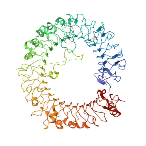
    6KYA

  • PubMed Abstract: 

    Rational designs of small-molecule inhibitors targeting protein-protein interfaces have met little success. Herein, we have designed a series of triazole derivatives with a novel scaffold to specifically intervene with the interaction of TLR8 homomerization. In multiple assays, TH1027 was identified as a highly potent and specific inhibitor of TLR8. A successful solution of the X-ray crystal structure of TLR8 in complex with TH1027 provided an in-depth mechanistic insight into its binding mode, validating that TH1027 was located between two TLR8 monomers and recognized as an unconventional pocket, thereby preventing TLR8 from activation. Further biological evaluations showed that TH1027 dose-dependently suppressed the TLR8-mediated inflammatory responses in both human monocyte cell lines, peripheral blood mononuclear cells, and rheumatoid arthritis patient specimens, suggesting a strong therapeutic potential against autoimmune diseases.


  • Organizational Affiliation
    • Graduate School of Pharmaceutical Sciences, The University of Tokyo, Tokyo 113-0033, Japan.

Macromolecule Content 

  • Total Structure Weight: 192.19 kDa 
  • Atom Count: 12,305 
  • Modeled Residue Count: 1,473 
  • Deposited Residue Count: 1,622 
  • Unique protein chains: 1

Macromolecules

Find similar proteins by:|  3D Structure
Entity ID: 1
MoleculeChains  Sequence LengthOrganismDetailsImage
Toll-like receptor 8
A, B
811Homo sapiensMutation(s): 0 
Gene Names: TLR8UNQ249/PRO286
UniProt & NIH Common Fund Data Resources
Find proteins for Q9NR97 (Homo sapiens)
Explore Q9NR97 
Go to UniProtKB:  Q9NR97
PHAROS:  Q9NR97
GTEx:  ENSG00000101916 
Entity Groups
Sequence Clusters30% Identity50% Identity70% Identity90% Identity95% Identity100% Identity
UniProt GroupQ9NR97
Glycosylation
Glycosylation Sites: 7Go to GlyGen: Q9NR97-1
Sequence Annotations
Expand
Reference Sequence

Oligosaccharides

Help  
Entity ID: 2
MoleculeChains Length2D Diagram GlycosylationD Interactions
alpha-D-mannopyranose-(1-3)-beta-D-mannopyranose-(1-4)-2-acetamido-2-deoxy-beta-D-glucopyranose-(1-4)-2-acetamido-2-deoxy-beta-D-glucopyranose
C, F
4N-Glycosylation
Glycosylation Resources
GlyTouCan: G81315DD
GlyCosmos: G81315DD
GlyGen: G81315DD
Entity ID: 3
MoleculeChains Length2D Diagram GlycosylationD Interactions
2-acetamido-2-deoxy-beta-D-glucopyranose-(1-4)-2-acetamido-2-deoxy-beta-D-glucopyranose
D, G
2N-Glycosylation
Glycosylation Resources
GlyTouCan: G42666HT
GlyCosmos: G42666HT
GlyGen: G42666HT
Entity ID: 4
MoleculeChains Length2D Diagram GlycosylationD Interactions
alpha-D-mannopyranose-(1-3)-[alpha-D-mannopyranose-(1-6)]beta-D-mannopyranose-(1-4)-2-acetamido-2-deoxy-beta-D-glucopyranose-(1-4)-2-acetamido-2-deoxy-beta-D-glucopyranose
E, H
5N-Glycosylation
Glycosylation Resources
GlyTouCan: G22768VO
GlyCosmos: G22768VO
GlyGen: G22768VO

Small Molecules

Ligands 2 Unique
IDChains Name / Formula / InChI Key2D Diagram3D Interactions
DY3
(Subject of Investigation/LOI)

Query on DY3


Download:Ideal Coordinates CCD File
I [auth A],
N [auth B]
3-[5-methylsulfanyl-4-(3,4,5-trimethylphenyl)-1,2,4-triazol-3-yl]propan-1-ol
C15 H21 N3 O S
WLYAKMVSMAFGQO-UHFFFAOYSA-N
NAG

Query on NAG


Download:Ideal Coordinates CCD File
J [auth A]
K [auth A]
L [auth A]
M [auth A]
O [auth B]
J [auth A],
K [auth A],
L [auth A],
M [auth A],
O [auth B],
P [auth B],
Q [auth B],
R [auth B]
2-acetamido-2-deoxy-beta-D-glucopyranose
C8 H15 N O6
OVRNDRQMDRJTHS-FMDGEEDCSA-N

Experimental Data & Validation

Experimental Data

  • Method: X-RAY DIFFRACTION
  • Resolution: 2.89 Å
  • R-Value Free:  0.277 (Depositor), 0.273 (DCC) 
  • R-Value Work:  0.228 (Depositor), 0.223 (DCC) 
  • R-Value Observed: 0.230 (Depositor) 
Space Group: C 1 2 1
Unit Cell:
Length ( Å )Angle ( ˚ )
a = 142.745α = 90
b = 96.871β = 107.65
c = 140.864γ = 90
Software Package:
Software NamePurpose
REFMACrefinement
PDB_EXTRACTdata extraction
HKL-2000data reduction
HKL-2000data scaling
MOLREPphasing

Structure Validation

View Full Validation Report


Ligand Structure Quality Assessment 


Entry History 

Deposition Data

Revision History  (Full details and data files)

  • Version 1.0: 2020-05-27
    Type: Initial release
  • Version 2.0: 2020-07-29
    Type: Remediation
    Reason: Carbohydrate remediation
    Changes: Atomic model, Data collection, Derived calculations, Structure summary
  • Version 2.1: 2023-11-22
    Changes: Data collection, Database references, Derived calculations, Refinement description, Structure summary
  • Version 2.2: 2024-11-06
    Changes: Structure summary