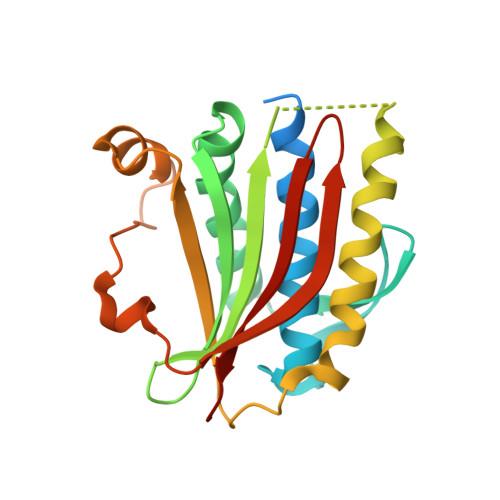
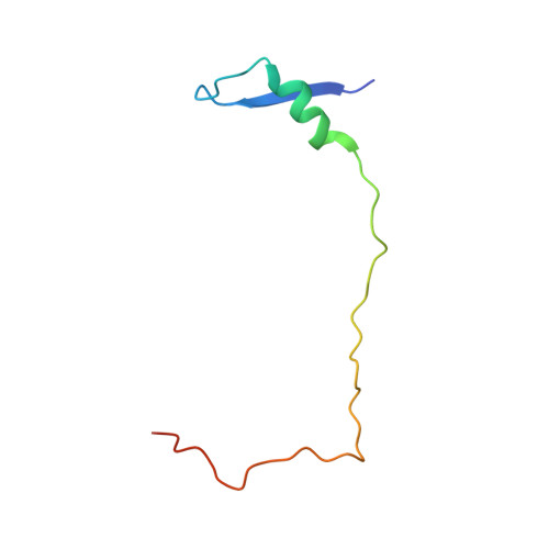
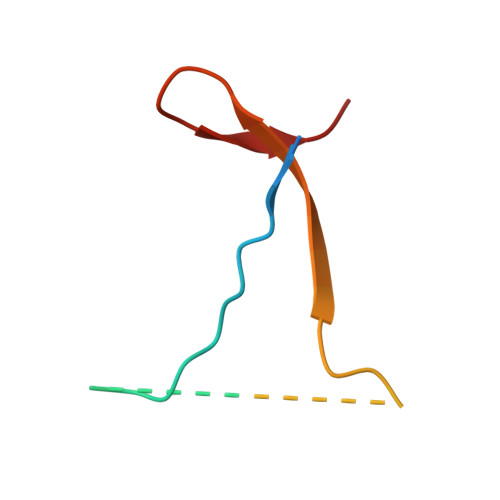
6KTO

Crystal structure of human SHLD3-C-REV7-O-REV7-SHLD2 complex


Experimental Data Snapshot

  • Method: X-RAY DIFFRACTION
  • Resolution: 3.45 Å
  • R-Value Free: 0.269 
  • R-Value Work: 0.242 
  • R-Value Observed: 0.243 

Starting Model: experimental
View more details

wwPDB Validation   3D Report Full Report


This is version 1.2 of the entry. See complete history


Literature

Molecular basis for assembly of the shieldin complex and its implications for NHEJ.

Liang, L.Feng, J.Zuo, P.Yang, J.Lu, Y.Yin, Y.

(2020) Nat Commun 11: 1972-1972

  • DOI: https://doi.org/10.1038/s41467-020-15879-5
  • Primary Citation of Related Structures:  
    6KTO

  • PubMed Abstract: 

    Shieldin, including SHLD1, SHLD2, SHLD3 and REV7, functions as a bridge linking 53BP1-RIF1 and single-strand DNA to suppress the DNA termini nucleolytic resection during non-homologous end joining (NHEJ). However, the mechanism of shieldin assembly remains unclear. Here we present the crystal structure of the SHLD3-REV7-SHLD2 ternary complex and reveal an unexpected C (closed)-REV7-O (open)-REV7 conformational dimer mediated by SHLD3. We show that SHLD2 interacts with O-REV7 and the N-terminus of SHLD3 by forming β sheet sandwich. Disruption of the REV7 conformational dimer abolishes the assembly of shieldin and impairs NHEJ efficiency. The conserved FXPWFP motif of SHLD3 binds to C-REV7 and blocks its binding to REV1, which excludes shieldin from the REV1/Pol ζ translesion synthesis (TLS) complex. Our study reveals the molecular architecture of shieldin assembly, elucidates the structural basis of the REV7 conformational dimer, and provides mechanistic insight into orchestration between TLS and NHEJ.


  • Organizational Affiliation

    Institute of Systems Biomedicine, Department of Pathology, Beijing Key Laboratory of Tumor Systems Biology, School of Basic Medical Sciences, Peking University Health Science Center, Beijing, 100191, China. liangling@hsc.pku.edu.cn.


Macromolecules
Find similar proteins by:  (by identity cutoff)  |  3D Structure
Entity ID: 1
MoleculeChains Sequence LengthOrganismDetailsImage
Mitotic spindle assembly checkpoint protein MAD2B
A, B
227Homo sapiensMutation(s): 0 
Gene Names: MAD2L2MAD2BREV7
UniProt & NIH Common Fund Data Resources
Find proteins for Q9UI95 (Homo sapiens)
Explore Q9UI95 
Go to UniProtKB:  Q9UI95
PHAROS:  Q9UI95
GTEx:  ENSG00000116670 
Entity Groups  
Sequence Clusters30% Identity50% Identity70% Identity90% Identity95% Identity100% Identity
UniProt GroupQ9UI95
Sequence Annotations
Expand
  • Reference Sequence
Find similar proteins by:  (by identity cutoff)  |  3D Structure
Entity ID: 2
MoleculeChains Sequence LengthOrganismDetailsImage
Shieldin complex subunit 364Homo sapiensMutation(s): 0 
Gene Names: SHLD3FLJ26957RINN1
UniProt & NIH Common Fund Data Resources
Find proteins for Q6ZNX1 (Homo sapiens)
Explore Q6ZNX1 
Go to UniProtKB:  Q6ZNX1
PHAROS:  Q6ZNX1
GTEx:  ENSG00000253251 
Entity Groups  
Sequence Clusters30% Identity50% Identity70% Identity90% Identity95% Identity100% Identity
UniProt GroupQ6ZNX1
Sequence Annotations
Expand
  • Reference Sequence
Find similar proteins by:  (by identity cutoff)  |  3D Structure
Entity ID: 3
MoleculeChains Sequence LengthOrganismDetailsImage
Shieldin complex subunit 254Homo sapiensMutation(s): 0 
Gene Names: SHLD2FAM35ARINN2
UniProt & NIH Common Fund Data Resources
Find proteins for Q86V20 (Homo sapiens)
Explore Q86V20 
Go to UniProtKB:  Q86V20
PHAROS:  Q86V20
GTEx:  ENSG00000122376 
Entity Groups  
Sequence Clusters30% Identity50% Identity70% Identity90% Identity95% Identity100% Identity
UniProt GroupQ86V20
Sequence Annotations
Expand
  • Reference Sequence
Experimental Data & Validation

Experimental Data

  • Method: X-RAY DIFFRACTION
  • Resolution: 3.45 Å
  • R-Value Free: 0.269 
  • R-Value Work: 0.242 
  • R-Value Observed: 0.243 
  • Space Group: P 61 2 2
Unit Cell:
Length ( Å )Angle ( ˚ )
a = 93.843α = 90
b = 93.843β = 90
c = 325.377γ = 120
Software Package:
Software NamePurpose
Cootmodel building
PHENIXrefinement
HKL-2000data reduction
HKL-2000data scaling
PHASERphasing

Structure Validation

View Full Validation Report


Entry History & Funding Information

Deposition Data


Funding OrganizationLocationGrant Number
National Natural Science Foundation of ChinaChina--

Revision History  (Full details and data files)

  • Version 1.0: 2020-04-29
    Type: Initial release
  • Version 1.1: 2020-05-06
    Changes: Database references
  • Version 1.2: 2023-11-22
    Changes: Data collection, Database references, Refinement description