6KRB | pdb_00006krb

High resolution crystal structure of an Acylphosphatase protein cage


Experimental Data Snapshot

  • Method: X-RAY DIFFRACTION
  • Resolution: 2.38 Å
  • R-Value Free: 
    0.241 (Depositor), 0.240 (DCC) 
  • R-Value Work: 
    0.194 (Depositor), 0.200 (DCC) 
  • R-Value Observed: 
    0.196 (Depositor) 

Starting Model: experimental
View more details

wwPDB Validation   3D Report Full Report


Ligand Structure Quality Assessment 


This is version 1.2 of the entry. See complete history


Literature

High resolution structure of Vibrio cholerae acylphosphatase (VcAcP) cage: Identification of drugs, location of its binding site and engineering to facilitate cage formation.

Chatterjee, S.Nath, S.Sen, U.

(2020) Biochem Biophys Res Commun 523: 348-353

  • DOI: https://doi.org/10.1016/j.bbrc.2019.12.060
  • Primary Citation of Related Structures:  
    6KRB

  • PubMed Abstract: 

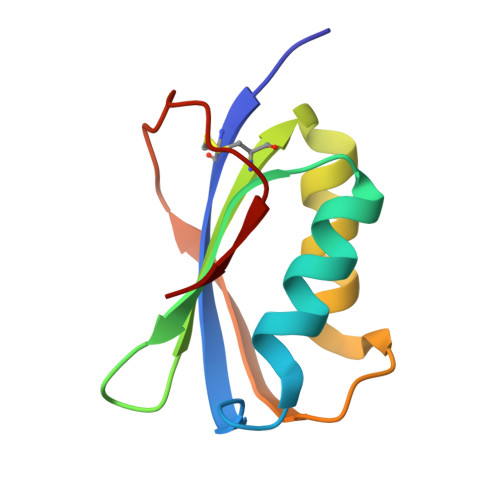
    Protein cages have recently emerged as an extraordinary drug-delivery system due to its biocompatibility, biodegradability, low toxicity, ease to manipulate and engineer. We have reported earlier the formation and architecture of a do-decameric cage-like architecture of Vibrio cholerae acylphosphatase (VcAcP) at 3.1 Å. High resolution (2.4 Å) crystal structure of VcAcP cage, reported here, illuminates a potential binding site for sulphate/phosphate containing drugs whereas analysis of its subunit association and interfaces indicates high potential for cage engineering. Tryptophan quenching studies indeed discloses noteworthy binding with various sulphate/phosphate containing nucleotide-based drugs and vitamin B 6 (PLP) demonstrating that exterior surface of VcAcP protein cage can be exploited as multifunctional carrier. Moreover, a quadruple mutant L30C/T68C/N40C/L81C-VcAcP (QM-VcAcP) capable to form an intricate disulphide bonded VcAcP cage has been designed. SEC, SDS-PAGE analysis and DLS experiment confirmed cysteine mediated engineered VcAcP cage formation.


  • Organizational Affiliation
    • Crystallography and Molecular Biology Division, Saha Institute of Nuclear Physics, HBNI, 1/AF Bidhan Nagar, Kolkata, 700064, India.

Macromolecules
Find similar proteins by:  (by identity cutoff)  |  3D Structure
Entity ID: 1
MoleculeChains Sequence LengthOrganismDetailsImage
Acylphosphatase
A, B, C, D, E
A, B, C, D, E, F, G, H, I, J, K, L
91Vibrio cholerae O395Mutation(s): 0 
Gene Names: acyPVC0395_A0969VC395_1474
EC: 3.6.1.7
UniProt
Find proteins for A5F8G9 (Vibrio cholerae serotype O1 (strain ATCC 39541 / Classical Ogawa 395 / O395))
Explore A5F8G9 
Go to UniProtKB:  A5F8G9
Entity Groups  
Sequence Clusters30% Identity50% Identity70% Identity90% Identity95% Identity100% Identity
UniProt GroupA5F8G9
Sequence Annotations
Expand
  • Reference Sequence
Small Molecules
Ligands 2 Unique
IDChains Name / Formula / InChI Key2D Diagram3D Interactions
SO4 (Subject of Investigation/LOI)
Query on SO4

Download Ideal Coordinates CCD File 
AA [auth C]
AB [auth H]
AC [auth L]
BA [auth C]
BB [auth H]
AA [auth C],
AB [auth H],
AC [auth L],
BA [auth C],
BB [auth H],
CB [auth H],
DA [auth D],
DB [auth H],
EA [auth D],
EB [auth H],
FA [auth D],
FB [auth I],
GA [auth D],
GB [auth I],
HA [auth D],
HB [auth I],
IA [auth E],
IB [auth I],
JA [auth E],
JB [auth I],
KA [auth E],
KB [auth I],
LA [auth E],
M [auth A],
MA [auth E],
N [auth A],
NA [auth F],
NB [auth J],
O [auth A],
OA [auth F],
OB [auth J],
P [auth A],
PA [auth F],
PB [auth J],
QA [auth F],
QB [auth J],
R [auth B],
S [auth B],
SB [auth K],
T [auth B],
TA [auth G],
TB [auth K],
U [auth B],
UA [auth G],
UB [auth K],
V [auth B],
VA [auth G],
VB [auth K],
W [auth B],
WA [auth G],
WB [auth K],
X [auth C],
XA [auth G],
XB [auth L],
Y [auth C],
YA [auth H],
YB [auth L],
Z [auth C],
ZA [auth H],
ZB [auth L]
SULFATE ION
O4 S
QAOWNCQODCNURD-UHFFFAOYSA-L
GOL
Query on GOL

Download Ideal Coordinates CCD File 
CA [auth C]
LB [auth I]
MB [auth I]
Q [auth A]
RA [auth F]
CA [auth C],
LB [auth I],
MB [auth I],
Q [auth A],
RA [auth F],
RB [auth J],
SA [auth F]
GLYCEROL
C3 H8 O3
PEDCQBHIVMGVHV-UHFFFAOYSA-N
Experimental Data & Validation

Experimental Data

  • Method: X-RAY DIFFRACTION
  • Resolution: 2.38 Å
  • R-Value Free:  0.241 (Depositor), 0.240 (DCC) 
  • R-Value Work:  0.194 (Depositor), 0.200 (DCC) 
  • R-Value Observed: 0.196 (Depositor) 
Space Group: P 31
Unit Cell:
Length ( Å )Angle ( ˚ )
a = 104.31α = 90
b = 104.31β = 90
c = 146.43γ = 120
Software Package:
Software NamePurpose
PHENIXrefinement
iMOSFLMdata reduction
Aimlessdata scaling
PHASERphasing

Structure Validation

View Full Validation Report


Ligand Structure Quality Assessment 


Entry History 

Deposition Data

Revision History  (Full details and data files)

  • Version 1.0: 2020-05-13
    Type: Initial release
  • Version 1.1: 2023-11-22
    Changes: Data collection, Database references, Refinement description
  • Version 1.2: 2024-11-13
    Changes: Structure summary