


Crystal structure of diamondback moth ryanodine receptor SPRY2 domain
Zhou, Y., Lin, L., Yuchi, Z.To be published.
Experimental Data Snapshot
Starting Model: experimental
View more details
Entity ID: 1 | |||||
|---|---|---|---|---|---|
| Molecule | Chains | Sequence Length | Organism | Details | Image |
| Ryanodine receptor | 180 | Plutella xylostella | Mutation(s): 1 | 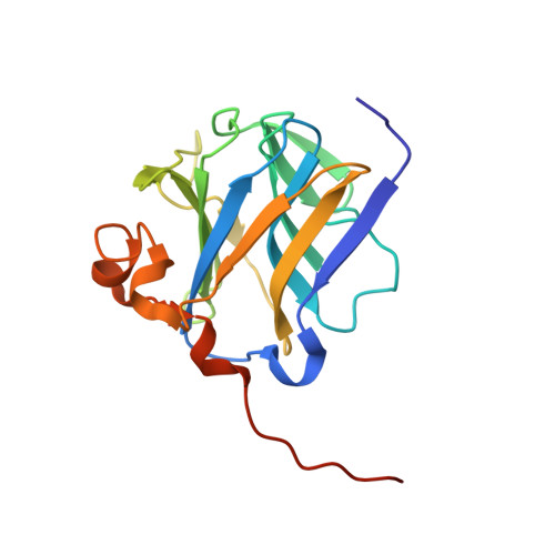 | |
UniProt | |||||
Entity Groups | |||||
| Sequence Clusters | 30% Identity50% Identity70% Identity90% Identity95% Identity100% Identity | ||||
| UniProt Group | I3VR32 | ||||
Sequence AnnotationsExpand | |||||
Reference Sequence | |||||
| Ligands 1 Unique | |||||
|---|---|---|---|---|---|
| ID | Chains | Name / Formula / InChI Key | 2D Diagram | 3D Interactions | |
| GOL (Subject of Investigation/LOI) Download:Ideal Coordinates CCD File | B [auth A], C [auth A], D [auth A], E [auth A] | GLYCEROL C3 H8 O3 PEDCQBHIVMGVHV-UHFFFAOYSA-N |  | ||
| Length ( Å ) | Angle ( ˚ ) |
|---|---|
| a = 68.025 | α = 90 |
| b = 37.637 | β = 93.24 |
| c = 67.476 | γ = 90 |
| Software Name | Purpose |
|---|---|
| SCALA | data scaling |
| PHENIX | refinement |
| PDB_EXTRACT | data extraction |
| HKL-3000 | data reduction |
| PHENIX | phasing |
| Funding Organization | Location | Grant Number |
|---|---|---|
| Ministry of Science and Technology (China) | China | 2017YFD0201400 |
| Ministry of Science and Technology (China) | China | 2017YFD0201403 |


