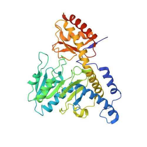
Snapshots of PLP-substrate and PLP-product external aldimines as intermediates in two types of cysteine desulfurase enzymes.
Nakamura, R., Hikita, M., Ogawa, S., Takahashi, Y., Fujishiro, T.(2020) FEBS J 287: 1138-1154
- PubMed: 31587510 Search on PubMed
- DOI: https://doi.org/10.1111/febs.15081
- Primary Citation Related Structures:
5WT2, 5WT4, 5ZS9, 5ZSK, 5ZSO, 5ZSP, 5ZST, 6KFZ, 6KG0, 6KG1 - PubMed Abstract:
Cysteine desulfurase enzymes catalyze sulfur mobilization from l-cysteine to sulfur-containing biomolecules such as iron-sulfur (Fe-S) clusters and thio-tRNAs. The enzymes utilize the cofactor pyridoxal-5'-phosphate (PLP), which forms the external substrate- and product-aldimines and ketimines during catalysis and are grouped into two types (I and II) based on their different catalytic loops. To clarify the structure-based catalytic mechanisms for each group, we determined the structures of the external substrate- and product-aldimines as catalytic intermediates of NifS (type I) and SufS (type II) that are involved in Fe-S cluster biosynthesis using X-ray crystallographic snapshot analysis. As a common intermediate structure, the thiol group of the PLP-l-cysteine external aldimine is stabilized by the conserved histidine adjacent to PLP through a polar interaction. This interaction makes the thiol group orientated for subsequent nucleophilic attack by a conserved cysteine residue on the catalytic loop in the state of PLP-l-cysteine ketimine, which is formed from the PLP-l-cysteine aldimine. Unlike the intermediates, structural changes of the loops were different between the type I and II enzymes. In the type I enzyme, conformational and topological change of the loop is necessary for nucleophilic attack by the cysteine. In contrast, the loop in type II cysteine desulfurase enzymes showed no large conformational change; rather, it might possibly orient the thiol group of the catalytic cysteine for nucleophilic attack toward PLP-l-cysteine. The present structures allow a revision of the catalytic mechanism and may provide a clue for consideration of enzyme function, structural diversity, and evolution of cysteine desulfurase enzymes. DATABASE: Structural data are available in PDB database under the accession numbers 5WT2, 5WT4, 5ZSP, 5ZST, 5ZS9, 5ZSK, 5ZSO, 6KFZ, 6KG0, and 6KG1.
- Department of Biochemistry and Molecular Biology, Graduate School of Science and Engineering, Saitama University, Saitama, Japan.
Organizational Affiliation: