6KCS

Crystal structure of HIRAN domain of HLTF in complex with duplex DNA


Experimental Data Snapshot

  • Method: X-RAY DIFFRACTION
  • Resolution: 2.10 Å
  • R-Value Free: 0.264 
  • R-Value Work: 0.229 
  • R-Value Observed: 0.230 

wwPDB Validation   3D Report Full Report


This is version 1.2 of the entry. See complete history


Literature

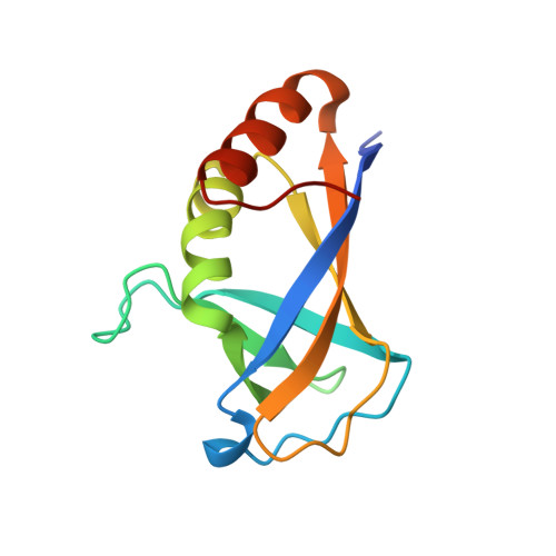
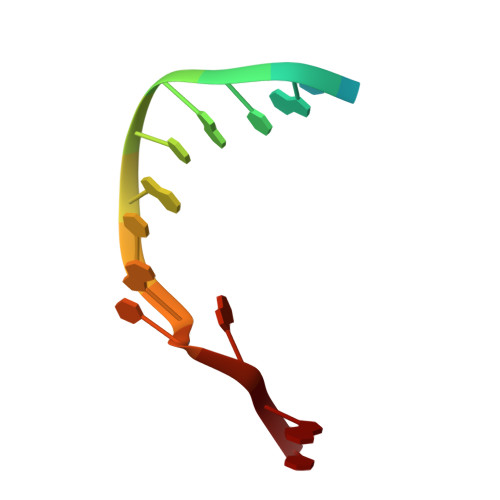
Structure of HIRAN domain of human HLTF bound to duplex DNA provides structural basis for DNA unwinding to initiate replication fork regression.

Hishiki, A.Sato, M.Hashimoto, H.

(2020) J Biochem 167: 597-602

  • DOI: https://doi.org/10.1093/jb/mvaa008
  • Primary Citation of Related Structures:  
    6KCS

  • PubMed Abstract: 

    Replication fork regression is a mechanism to rescue a stalled fork by various replication stresses, such as DNA lesions. Helicase-like transcription factor, a SNF2 translocase, plays a central role in the fork regression and its N-terminal domain, HIRAN (HIP116 and Rad5 N-terminal), binds the 3'-hydroxy group of single-stranded DNA. Furthermore, HIRAN is supposed to bind double-stranded DNA (dsDNA) and involved in strand separation in the fork regression, whereas structural basis for mechanisms underlying dsDNA binding and strand separation by HIRAN are still unclear. Here, we report the crystal structure of HIRAN bound to duplex DNA. The structure reveals that HIRAN binds the 3'-hydroxy group of DNA and unexpectedly unwinds three nucleobases of the duplex. Phe-142 is involved in the dsDNA binding and the strand separation. In addition, the structure unravels the mechanism underlying sequence-independent recognition for purine bases by HIRAN, where the N-glycosidic bond adopts syn conformation. Our findings indicate direct involvement of HIRAN in the fork regression by separating of the daughter strand from the parental template.


  • Organizational Affiliation

    School of Pharmaceutical Sciences, University of Shizuoka, 52-1 Yada, Suruga-ku, Shizuoka 422-8002, Japan.


Macromolecules

Find similar proteins by:  (by identity cutoff)  |  3D Structure
Entity ID: 1
MoleculeChains Sequence LengthOrganismDetailsImage
Helicase-like transcription factor122Homo sapiensMutation(s): 0 
Gene Names: HLTFHIP116ARNF80SMARCA3SNF2L3ZBU1
EC: 2.3.2.27 (PDB Primary Data), 3.6.4 (PDB Primary Data)
UniProt & NIH Common Fund Data Resources
Find proteins for Q14527 (Homo sapiens)
Explore Q14527 
Go to UniProtKB:  Q14527
PHAROS:  Q14527
GTEx:  ENSG00000071794 
Entity Groups  
Sequence Clusters30% Identity50% Identity70% Identity90% Identity95% Identity100% Identity
UniProt GroupQ14527
Sequence Annotations
Expand
  • Reference Sequence

Find similar nucleic acids by:  Sequence   |   3D Structure  

Entity ID: 2
MoleculeChains LengthOrganismImage
DNA (5'-D(*AP*CP*TP*GP*TP*AP*CP*GP*TP*AP*CP*AP*GP*T)-3')14synthetic construct
Sequence Annotations
Expand
  • Reference Sequence
Experimental Data & Validation

Experimental Data

  • Method: X-RAY DIFFRACTION
  • Resolution: 2.10 Å
  • R-Value Free: 0.264 
  • R-Value Work: 0.229 
  • R-Value Observed: 0.230 
  • Space Group: P 31 2 1
Unit Cell:
Length ( Å )Angle ( ˚ )
a = 45.363α = 90
b = 45.363β = 90
c = 125.827γ = 120
Software Package:
Software NamePurpose
REFMACrefinement
XDSdata reduction
SCALAdata scaling
MOLREPphasing

Structure Validation

View Full Validation Report


Entry History & Funding Information

Deposition Data


Funding OrganizationLocationGrant Number
Japan Society for the Promotion of ScienceJapan25291017
Japan Society for the Promotion of ScienceJapan16H04755
Japan Society for the Promotion of ScienceJapan23770125

Revision History  (Full details and data files)

  • Version 1.0: 2020-06-10
    Type: Initial release
  • Version 1.1: 2020-12-23
    Changes: Derived calculations
  • Version 1.2: 2023-11-22
    Changes: Data collection, Database references, Refinement description