


Crystal structure of ErbB3 N418Q mutant
Takahashi, M., Kato, K., Yao, M.To be published.
Experimental Data Snapshot
Starting Model: experimental
View more details
Entity ID: 1 | |||||
|---|---|---|---|---|---|
| Molecule | Chains | Sequence Length | Organism | Details | Image |
| Receptor tyrosine-protein kinase erbB-3 | 620 | Homo sapiens | Mutation(s): 1 Gene Names: ERBB3, HER3 EC: 2.7.10.1 | 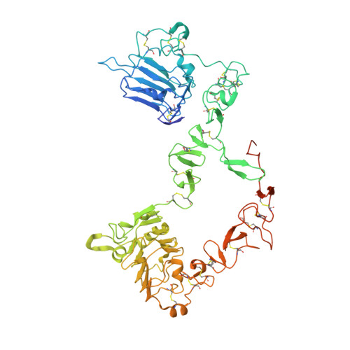 | |
UniProt & NIH Common Fund Data Resources | |||||
PHAROS: P21860 GTEx: ENSG00000065361 | |||||
Entity Groups | |||||
| Sequence Clusters | 30% Identity50% Identity70% Identity90% Identity95% Identity100% Identity | ||||
| UniProt Group | P21860 | ||||
Glycosylation | |||||
| Glycosylation Sites: 8 | Go to GlyGen: P21860-1 | ||||
Sequence AnnotationsExpand | |||||
Reference Sequence | |||||
| Ligands 1 Unique | |||||
|---|---|---|---|---|---|
| ID | Chains | Name / Formula / InChI Key | 2D Diagram | 3D Interactions | |
| NAG Download:Ideal Coordinates CCD File | E [auth A] F [auth A] G [auth A] H [auth A] I [auth A] | 2-acetamido-2-deoxy-beta-D-glucopyranose C8 H15 N O6 OVRNDRQMDRJTHS-FMDGEEDCSA-N |  | ||
| Length ( Å ) | Angle ( ˚ ) |
|---|---|
| a = 236.105 | α = 90 |
| b = 49.458 | β = 125.62 |
| c = 190.919 | γ = 90 |
| Software Name | Purpose |
|---|---|
| PHENIX | refinement |
| XDS | data reduction |
| XDS | data scaling |
| PHENIX | phasing |


