6K9Y | pdb_00006k9y

Crystal structure of human VAT-1


Experimental Data Snapshot

  • Method: X-RAY DIFFRACTION
  • Resolution: 2.20 Å
  • R-Value Free: 
    0.255 (Depositor), 0.254 (DCC) 
  • R-Value Work: 
    0.222 (Depositor), 0.222 (DCC) 

Starting Model: experimental
View more details

wwPDB Validation 3D Report Full Report

Validation slider image for 6K9Y

This is version 1.2 of the entry. See complete history

Literature

Structural basis for interorganelle phospholipid transport mediated by VAT-1.

Watanabe, Y.Tamura, Y.Kakuta, C.Watanabe, S.Endo, T.

(2020) J Biological Chem 295: 3257-3268

  • DOI: https://doi.org/10.1074/jbc.RA119.011019
  • Primary Citation Related Structures: 
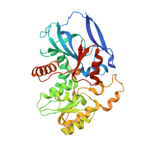
    6K9Y

  • PubMed Abstract: 

    Eukaryotic cells are compartmentalized to form organelles, whose functions rely on proper phospholipid and protein transport. Here we determined the crystal structure of human VAT-1, a cytosolic soluble protein that was suggested to transfer phosphatidylserine, at 2.2 Å resolution. We found that VAT-1 transferred not only phosphatidylserine but also other acidic phospholipids between membranes in vitro Structure-based mutational analyses showed the presence of a possible lipid-binding cavity at the interface between the two subdomains, and two tyrosine residues in the flexible loops facilitated phospholipid transfer, likely by functioning as a gate to this lipid-binding cavity. We also found that a basic and hydrophobic loop with two tryptophan residues protruded from the molecule and facilitated binding to the acidic-lipid membranes, thereby achieving efficient phospholipid transfer.


  • Organizational Affiliation
    • Department of Bioscience, Graduate School of Agriculture, Ehime University, Matsuyama, Ehime 790-8566, Japan; Faculty of Life Sciences, Kyoto Sangyo University, Kamigamo-Motoyama, Kita-ku, Kyoto 603-8555, Japan.

Macromolecule Content 

  • Total Structure Weight: 154.36 kDa 
  • Atom Count: 10,175 
  • Modeled Residue Count: 1,328 
  • Deposited Residue Count: 1,428 
  • Unique protein chains: 1

Macromolecules

Find similar proteins by:|  3D Structure
Entity ID: 1
MoleculeChains  Sequence LengthOrganismDetailsImage
Synaptic vesicle membrane protein VAT-1 homolog
A, B, C, D
357Homo sapiensMutation(s): 0 
Gene Names: VAT1
EC: 1 (PDB Primary Data), 1.6.5.5 (UniProt)
UniProt & NIH Common Fund Data Resources
Find proteins for Q99536 (Homo sapiens)
Explore Q99536 
Go to UniProtKB:  Q99536
PHAROS:  Q99536
GTEx:  ENSG00000108828 
Entity Groups
Sequence Clusters30% Identity50% Identity70% Identity90% Identity95% Identity100% Identity
UniProt GroupQ99536
Sequence Annotations
Expand
Reference Sequence

Experimental Data & Validation

Experimental Data

  • Method: X-RAY DIFFRACTION
  • Resolution: 2.20 Å
  • R-Value Free:  0.255 (Depositor), 0.254 (DCC) 
  • R-Value Work:  0.222 (Depositor), 0.222 (DCC) 
Space Group: P 21 21 21
Unit Cell:
Length ( Å )Angle ( ˚ )
a = 67.155α = 90
b = 131.72β = 90
c = 174.027γ = 90
Software Package:
Software NamePurpose
XSCALEdata scaling
CNSrefinement
PDB_EXTRACTdata extraction
PHASERphasing

Structure Validation

View Full Validation Report


Entry History 

Deposition Data

Revision History  (Full details and data files)

  • Version 1.0: 2020-02-12
    Type: Initial release
  • Version 1.1: 2020-03-18
    Changes: Database references
  • Version 1.2: 2023-11-22
    Changes: Data collection, Database references, Refinement description