6K8Z | pdb_00006k8z

Pyridoxal Kinase from Leishmania donovani in complex with ADP


Experimental Data Snapshot

  • Method: X-RAY DIFFRACTION
  • Resolution: 1.90 Å
  • R-Value Free: 
    0.217 (Depositor), 0.220 (DCC) 
  • R-Value Work: 
    0.173 (Depositor), 0.180 (DCC) 
  • R-Value Observed: 
    0.175 (Depositor) 

Starting Model: experimental
View more details

wwPDB Validation 3D Report Full Report

Validation slider image for 6K8Z

Ligand Structure Quality Assessment 


This is version 1.1 of the entry. See complete history

Literature

Structural attributes and substrate specificity of pyridoxal kinase from Leishmania donovani.

Are, S.Gatreddi, S.Jakkula, P.Qureshi, I.A.

(2020) Int J Biol Macromol 152: 812-827

  • DOI: https://doi.org/10.1016/j.ijbiomac.2020.02.257
  • Primary Citation Related Structures: 
    6K8Z, 6K90, 6K91, 6K92

  • PubMed Abstract: 

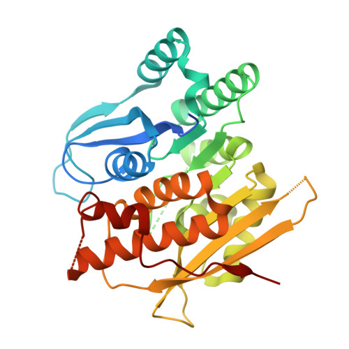
    The enzyme pyridoxal kinase (PdxK) catalyzes the conversion of pyridoxal to pyridoxal-5'-phosphate (PLP) using ATP as the co-factor. The product pyridoxal-5'-phosphate plays a key role in several biological processes such as transamination, decarboxylation and deamination. In the present study, full-length ORF of PdxK from Leishmania donovani (LdPdxK) was cloned and then purified using affinity chromatography. LdPdxK exists as a homo-dimer in solution and shows more activity at near to physiological pH. Biochemical analysis of LdPdxK with pyridoxal, pyridoxamine, pyridoxine and ginkgotoxin revealed its affinity preference towards different substrates. The secondary structure analysis using circular dichroism spectroscopy showed LdPdxK to be predominantly α-helical in organization which tends to decline at lower and higher pH. Simultaneously, LdPdxK was crystallized and its three-dimensional structure in complex with ADP and different substrates were determined. Crystal structure of LdPdxK delineated that it has a central core of β-sheets surrounded by α-helices with a conserved GTGD ribokinase motif. The structures of LdPdxK disclosed no major structural changes between ADP and ADP- substrate bound structures. In addition, comparative structural analysis highlighted the key differences between the active site pockets of leishmanial and human PdxK, rendering LdPdxK an attractive candidate for the designing of novel and specific inhibitors.


  • Organizational Affiliation
    • Department of Biotechnology and Bioinformatics, School of Life Sciences, University of Hyderabad, Hyderabad 500 046, Telangana, India.

Macromolecule Content 

  • Total Structure Weight: 71.93 kDa 
  • Atom Count: 4,596 
  • Modeled Residue Count: 580 
  • Deposited Residue Count: 644 
  • Unique protein chains: 1

Macromolecules

Find similar proteins by:|  3D Structure
Entity ID: 1
MoleculeChains  Sequence LengthOrganismDetailsImage
Pyridoxal kinase, putative
A, B
322Leishmania donovaniMutation(s): 0 
Gene Names: LdCL_300018300
EC: 2.7.1.35
UniProt
Find proteins for A0A3S7X3C0 (Leishmania donovani)
Explore A0A3S7X3C0 
Go to UniProtKB:  A0A3S7X3C0
Entity Groups
Sequence Clusters30% Identity50% Identity70% Identity90% Identity95% Identity100% Identity
UniProt GroupA0A3S7X3C0
Sequence Annotations
Expand
Reference Sequence

Small Molecules

Ligands 4 Unique
IDChains Name / Formula / InChI Key2D Diagram3D Interactions
ADP
(Subject of Investigation/LOI)

Query on ADP


Download:Ideal Coordinates CCD File
C [auth A],
H [auth B]
ADENOSINE-5'-DIPHOSPHATE
C10 H15 N5 O10 P2
XTWYTFMLZFPYCI-KQYNXXCUSA-N
ACT
(Subject of Investigation/LOI)

Query on ACT


Download:Ideal Coordinates CCD File
F [auth A],
M [auth B]
ACETATE ION
C2 H3 O2
QTBSBXVTEAMEQO-UHFFFAOYSA-M
CA
(Subject of Investigation/LOI)

Query on CA


Download:Ideal Coordinates CCD File
D [auth A]
E [auth A]
I [auth B]
J [auth B]
K [auth B]
D [auth A],
E [auth A],
I [auth B],
J [auth B],
K [auth B],
L [auth B]
CALCIUM ION
Ca
BHPQYMZQTOCNFJ-UHFFFAOYSA-N
CL

Query on CL


Download:Ideal Coordinates CCD File
G [auth A]CHLORIDE ION
Cl
VEXZGXHMUGYJMC-UHFFFAOYSA-M

Experimental Data & Validation

Experimental Data

  • Method: X-RAY DIFFRACTION
  • Resolution: 1.90 Å
  • R-Value Free:  0.217 (Depositor), 0.220 (DCC) 
  • R-Value Work:  0.173 (Depositor), 0.180 (DCC) 
  • R-Value Observed: 0.175 (Depositor) 
Space Group: P 1 21 1
Unit Cell:
Length ( Å )Angle ( ˚ )
a = 54.63α = 90
b = 71.08β = 98.93
c = 77.31γ = 90
Software Package:
Software NamePurpose
REFMACrefinement
iMOSFLMdata reduction
SCALAdata scaling
PHASERphasing

Structure Validation

View Full Validation Report


Ligand Structure Quality Assessment 


Entry History 

Deposition Data

Revision History  (Full details and data files)

  • Version 1.0: 2020-03-25
    Type: Initial release
  • Version 1.1: 2023-11-22
    Changes: Data collection, Database references, Derived calculations, Refinement description