


Crystal structure of glutathione S-transferase complexed and modified with glutathione
Okajima, T., Arai, R.To be published.
Experimental Data Snapshot
Starting Model: experimental
View more details
Entity ID: 1 | |||||
|---|---|---|---|---|---|
| Molecule | Chains | Sequence Length | Organism | Details | Image |
| Glutathione S-transferase class-mu 26 kDa isozyme | 226 | Schistosoma japonicum | Mutation(s): 0 EC: 2.5.1.18 | 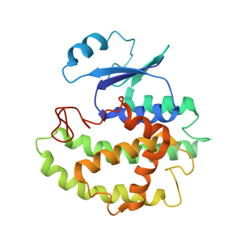 | |
UniProt | |||||
Entity Groups | |||||
| Sequence Clusters | 30% Identity50% Identity70% Identity90% Identity95% Identity100% Identity | ||||
| UniProt Group | P08515 | ||||
Sequence AnnotationsExpand | |||||
Reference Sequence | |||||
| Ligands 3 Unique | |||||
|---|---|---|---|---|---|
| ID | Chains | Name / Formula / InChI Key | 2D Diagram | 3D Interactions | |
| GSH Download:Ideal Coordinates CCD File | B [auth A], C [auth A], D [auth A], E [auth A] | Glutathione C10 H17 N3 O6 S RWSXRVCMGQZWBV-WDSKDSINSA-N |  | ||
| TRS Download:Ideal Coordinates CCD File | H [auth A] | 2-AMINO-2-HYDROXYMETHYL-PROPANE-1,3-DIOL C4 H12 N O3 LENZDBCJOHFCAS-UHFFFAOYSA-O |  | ||
| ACT Download:Ideal Coordinates CCD File | F [auth A], G [auth A] | ACETATE ION C2 H3 O2 QTBSBXVTEAMEQO-UHFFFAOYSA-M |  | ||
| Entity ID: 2 | |||||
|---|---|---|---|---|---|
| ID | Chains | Name | Type/Class | 2D Diagram | 3D Interactions |
| PRD_002593 (GSH) Query on PRD_002593 | B [auth A], C [auth A], D [auth A], E [auth A] | Glutathione | Peptide-like / Oxidation-reduction | | |
| Length ( Å ) | Angle ( ˚ ) |
|---|---|
| a = 93.708 | α = 90 |
| b = 93.708 | β = 90 |
| c = 57.609 | γ = 90 |
| Software Name | Purpose |
|---|---|
| REFMAC | refinement |
| HKL-2000 | data reduction |
| HKL-2000 | data scaling |
| PHASER | phasing |
| Funding Organization | Location | Grant Number |
|---|---|---|
| Japan Society for the Promotion of Science | Japan | 16K05841 |
| Japan Society for the Promotion of Science | Japan | 16H00761 |


