6JBM

Crystal structure of the TRIM14 PRYSPRY domain


Experimental Data Snapshot

  • Method: X-RAY DIFFRACTION
  • Resolution: 2.10 Å
  • R-Value Free: 0.242 
  • R-Value Work: 0.213 
  • R-Value Observed: 0.215 

wwPDB Validation   3D Report Full Report


This is version 1.1 of the entry. See complete history


Literature

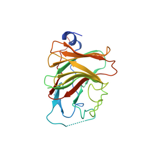
The TRIM14 PRYSPRY domain mediates protein interaction via its basic interface.

Yu, Y.Liang, L.Jin, Y.Yin, Y.

(2019) FEBS Lett 593: 1122-1129

  • DOI: https://doi.org/10.1002/1873-3468.13386
  • Primary Citation of Related Structures:  
    6JBM

  • PubMed Abstract: 

    Tripartite motif (TRIM)14 was recently shown to be an important molecule against pathogens. Its PRYSPRY domain acts as a protein-protein interaction module. TRIM14 exerts distinct functions via its PRYSPRY domain by interacting with different partners. However, the structural basis for its binding specificity remains unknown. Here we solved the crystal structure of the TRIM14 PRYSPRY domain, and found a positively charged surface that may mediate its partner specificity. Isothermal titration calorimetry reveals that the TRIM14 PRYSPRY domain binds to acidic peptides, and the analysis of the reported partners of TRIM14 is consistent with our assumption. Therefore, we demonstrate that the PRYSPRY domain of TRIM14 harbors a putative basic interface that may favorably bind to acidic amino acid residues.


  • Organizational Affiliation

    Institute of Systems Biomedicine, Department of Pathology, School of Basic Medical Sciences, Peking University Health Science Center, Beijing, China.


Macromolecules
Find similar proteins by:  (by identity cutoff)  |  3D Structure
Entity ID: 1
MoleculeChains Sequence LengthOrganismDetailsImage
Tripartite motif-containing protein 14A [auth B],
B [auth A],
C,
D
200Homo sapiensMutation(s): 0 
Gene Names: TRIM14KIAA0129
UniProt & NIH Common Fund Data Resources
Find proteins for Q14142 (Homo sapiens)
Explore Q14142 
Go to UniProtKB:  Q14142
PHAROS:  Q14142
GTEx:  ENSG00000106785 
Entity Groups  
Sequence Clusters30% Identity50% Identity70% Identity90% Identity95% Identity100% Identity
UniProt GroupQ14142
Sequence Annotations
Expand
  • Reference Sequence
Small Molecules
Ligands 1 Unique
IDChains Name / Formula / InChI Key2D Diagram3D Interactions
SO4
Query on SO4

Download Ideal Coordinates CCD File 
E [auth B]
F [auth B]
G [auth B]
H [auth B]
I [auth A]
E [auth B],
F [auth B],
G [auth B],
H [auth B],
I [auth A],
J [auth A],
K [auth A],
L [auth C],
M [auth D],
N [auth D],
O [auth D],
P [auth D]
SULFATE ION
O4 S
QAOWNCQODCNURD-UHFFFAOYSA-L
Experimental Data & Validation

Experimental Data

  • Method: X-RAY DIFFRACTION
  • Resolution: 2.10 Å
  • R-Value Free: 0.242 
  • R-Value Work: 0.213 
  • R-Value Observed: 0.215 
  • Space Group: P 1 21 1
Unit Cell:
Length ( Å )Angle ( ˚ )
a = 48.326α = 90
b = 104.02β = 91.49
c = 93.381γ = 90
Software Package:
Software NamePurpose
PHENIXrefinement
Cootmodel building
HKL-2000data scaling

Structure Validation

View Full Validation Report


Entry History & Funding Information

Deposition Data


Funding OrganizationLocationGrant Number
National Natural Science Foundation of ChinaChina81430056
National Natural Science Foundation of ChinaChina31420103905
National Natural Science Foundation of ChinaChina31800626

Revision History  (Full details and data files)

  • Version 1.0: 2020-03-04
    Type: Initial release
  • Version 1.1: 2023-11-22
    Changes: Data collection, Database references, Refinement description