6IYH

X-ray sequence and high resolution crystal structure of Persian sturgeon methemoglobin


Experimental Data Snapshot

  • Method: X-RAY DIFFRACTION
  • Resolution: 1.70 Å
  • R-Value Free: 0.196 
  • R-Value Work: 0.158 
  • R-Value Observed: 0.160 

wwPDB Validation   3D Report Full Report


Ligand Structure Quality Assessment 


This is version 1.1 of the entry. See complete history


Literature

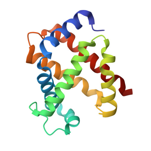
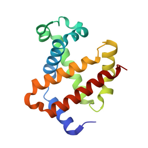
Novel X-ray sequences and crystal structures of Persian and Starry sturgeon methemoglobins: Highlighting the role of heme pocket waters in causing autoxidation.

Seyedarabi, A.Ariaeenejad, S.Moosavi-Movahedi, A.A.Rayati, S.Poursasan, N.Asiaie, N.Seraj, Z.Mehraban, F.Seyedarabi, S.E.

(2019) Biochim Biophys Acta Proteins Proteom 1867: 586-594

  • DOI: https://doi.org/10.1016/j.bbapap.2019.03.008
  • Primary Citation of Related Structures:  
    6IYH, 6IYI

  • PubMed Abstract: 
  • Although there is a high sequence similarity between mammalian and fish hemoglobin (Hb), the oxidation and heme loss rates can vary greatly between them such that fish Hbs oxidise much more rapidly than mammalian Hbs. There is to date no sequence or structural data for any sturgeon Hb to reveal the level of autoxidation in these fish ...

    Although there is a high sequence similarity between mammalian and fish hemoglobin (Hb), the oxidation and heme loss rates can vary greatly between them such that fish Hbs oxidise much more rapidly than mammalian Hbs. There is to date no sequence or structural data for any sturgeon Hb to reveal the level of autoxidation in these fish. In this study, novel high resolution X-ray sequences and crystal structures of methemoglobin (Met-Hb) from two sturgeon fish including Persian sturgeon (Acipenser percisus) and Starry sturgeon (Acipenser stellatus) belonging to the Caspian sea has been determined. A comprehensive sequence and structure comparison between these sturgeon Met-Hbs and a number of non-sturgeon and normal and sickle cell anaemia human Hb in varying heme states has been carried out highlighting (i) the structural variability in the heme propionate groups; (ii) the existence of certain residues or their displacement and shift in the heme pocket allowing entry of water molecules into the heme pocket; (iii) the importance of the number of water molecules in the heme pocket; (iv) the hydrogen bonding between oxygens of A and D propionate groups and that of waters in the heme pocket; and (v) the role of heme binding waters causing oxidative stress and heme autoxidation.


    Organizational Affiliation

    Institute of Biochemistry and Biophysics, University of Tehran, P.O. Box 13145-1384, Tehran, Iran.


Macromolecules
Find similar proteins by:  (by identity cutoff)  |  3D Structure
Entity ID: 1
MoleculeChainsSequence LengthOrganismDetailsImage
Alpha chain142Acipenser persicusMutation(s): 0 
UniProt
Find proteins for A0A1X8XL85 (Acipenser persicus)
Explore A0A1X8XL85 
Go to UniProtKB:  A0A1X8XL85
Entity Groups  
Sequence Clusters30% Identity50% Identity70% Identity90% Identity95% Identity100% Identity
UniProt GroupA0A1X8XL85
Protein Feature View
Expand
  • Reference Sequence
Find similar proteins by:  (by identity cutoff)  |  3D Structure
Entity ID: 2
MoleculeChainsSequence LengthOrganismDetailsImage
Beta chain145Acipenser persicusMutation(s): 0 
UniProt
Find proteins for A0A1X8XL86 (Acipenser persicus)
Explore A0A1X8XL86 
Go to UniProtKB:  A0A1X8XL86
Entity Groups  
Sequence Clusters30% Identity50% Identity70% Identity90% Identity95% Identity100% Identity
UniProt GroupA0A1X8XL86
Protein Feature View
Expand
  • Reference Sequence
Small Molecules
Ligands 2 Unique
IDChainsName / Formula / InChI Key2D Diagram3D Interactions
HEM
Query on HEM

Download Ideal Coordinates CCD File 
C [auth A],
D [auth B]
PROTOPORPHYRIN IX CONTAINING FE
C34 H32 Fe N4 O4
KABFMIBPWCXCRK-RGGAHWMASA-L
 Ligand Interaction
PGO
Query on PGO

Download Ideal Coordinates CCD File 
E [auth B]S-1,2-PROPANEDIOL
C3 H8 O2
DNIAPMSPPWPWGF-VKHMYHEASA-N
 Ligand Interaction
Experimental Data & Validation

Experimental Data

  • Method: X-RAY DIFFRACTION
  • Resolution: 1.70 Å
  • R-Value Free: 0.196 
  • R-Value Work: 0.158 
  • R-Value Observed: 0.160 
  • Space Group: I 1 2 1
Unit Cell:
Length ( Å )Angle ( ˚ )
a = 53.89α = 90
b = 62.29β = 97.67
c = 101.564γ = 90
Software Package:
Software NamePurpose
REFMACrefinement
iMOSFLMdata reduction
SCALAdata scaling
MOLREPphasing

Structure Validation

View Full Validation Report


Ligand Structure Quality Assessment 


Entry History 

Deposition Data

  • Released Date: 2019-04-10 
  • Deposition Author(s): Seyedarabi, A.
  • This entry supersedes: 5JGG

Revision History  (Full details and data files)

  • Version 1.0: 2019-04-10
    Type: Initial release
  • Version 1.1: 2019-04-17
    Changes: Data collection, Database references