6IWV

Crystal structure of a single strand DNA binding protein


Experimental Data Snapshot

  • Method: X-RAY DIFFRACTION
  • Resolution: 1.52 Å
  • R-Value Free: 0.181 
  • R-Value Work: 0.146 
  • R-Value Observed: 0.147 

wwPDB Validation   3D Report Full Report


This is version 1.1 of the entry. See complete history


Literature

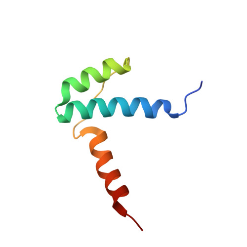
Crystal structure of the LUFS domain of human single-stranded DNA binding Protein 2 (SSBP2).

Wang, H.Wang, Z.Tang, Q.Yan, X.X.Xu, W.

(2019) Protein Sci 28: 788-793

  • DOI: https://doi.org/10.1002/pro.3581
  • Primary Citation of Related Structures:  
    6IWV

  • PubMed Abstract: 

    The human single-stranded DNA binding Protein 2 (SSBP2) is a tumor suppressor implicated in multiple cancer forms. The SSBP2 and related SSBP3/SSBP4 proteins are predicted to be intrinsically disordered excepted for their highly conserved N-terminal LUFS (LUG/LUH, Flo8, and SSBP/SSDP) domain. LUFS domains are found in a number of proteins including some transcriptional co-repressors. Although LUFS domains contain an N-terminal Lis homology (LisH) motif that typically forms a stable dimer, no 3D structure of any LUFS domain is available. Here, we report a crystal structure of the LUFS domain of human SSBP2 at 1.52 Å resolution. We show that the SSBP2 LUFS domain forms a homo-tetramer and reveal how an alpha-helix C-terminal to the LisH motif mediates SSBP2 tetramerization (dimerization of dimers). Conservation of the tetramerization interface among LUFS domains suggests that other LUFS domains may also form tetramers in similar manners.


  • Organizational Affiliation

    National Laboratory of Biomacromolecules, CAS Center for Excellence in Biomacromolecules, Institute of Biophysics, Chinese Academy of Sciences, Beijing 100101, People's Republic of China.


Macromolecules
Find similar proteins by:  (by identity cutoff)  |  3D Structure
Entity ID: 1
MoleculeChains Sequence LengthOrganismDetailsImage
Single-stranded DNA-binding protein 2
A, B
68Homo sapiensMutation(s): 0 
Gene Names: SSBP2SSDP2
UniProt & NIH Common Fund Data Resources
Find proteins for P81877 (Homo sapiens)
Explore P81877 
Go to UniProtKB:  P81877
PHAROS:  P81877
GTEx:  ENSG00000145687 
Entity Groups  
Sequence Clusters30% Identity50% Identity70% Identity90% Identity95% Identity100% Identity
UniProt GroupP81877
Sequence Annotations
Expand
  • Reference Sequence
Experimental Data & Validation

Experimental Data

  • Method: X-RAY DIFFRACTION
  • Resolution: 1.52 Å
  • R-Value Free: 0.181 
  • R-Value Work: 0.146 
  • R-Value Observed: 0.147 
  • Space Group: P 63 2 2
Unit Cell:
Length ( Å )Angle ( ˚ )
a = 116.906α = 90
b = 116.906β = 90
c = 42.355γ = 120
Software Package:
Software NamePurpose
REFMACrefinement
HKL-3000data collection
HKL-3000data scaling
AutoSolphasing
HKL-3000data reduction

Structure Validation

View Full Validation Report


Entry History 

Deposition Data

Revision History  (Full details and data files)

  • Version 1.0: 2019-08-14
    Type: Initial release
  • Version 1.1: 2024-03-27
    Changes: Data collection, Database references