6ISS

Lignin peroxidase H8 triple mutant S49C/A67C/H239


Experimental Data Snapshot

  • Method: X-RAY DIFFRACTION
  • Resolution: 1.53 Å
  • R-Value Free: 0.196 
  • R-Value Work: 0.170 
  • R-Value Observed: 0.171 

wwPDB Validation   3D Report Full Report


Ligand Structure Quality Assessment 


This is version 1.2 of the entry. See complete history


Literature

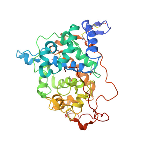
Extra disulfide and ionic salt bridge improves the thermostability of lignin peroxidase H8 under acidic condition

Son, H.Seo, H.Han, S.Kim, S.Thanh, L.Khan, M.Sung, H.Kang, S.H.Kim, K.J.Kim, Y.

(2021) Enzyme Microb Technol 148: 109803


Macromolecules
Find similar proteins by:  (by identity cutoff)  |  3D Structure
Entity ID: 1
MoleculeChains Sequence LengthOrganismDetailsImage
Ligninase H8A,
B [auth G]
351Phanerochaete chrysosporium RP-78Mutation(s): 4 
Gene Names: LPOA
EC: 1.11.1.14
UniProt
Find proteins for P06181 (Phanerodontia chrysosporium)
Explore P06181 
Go to UniProtKB:  P06181
Entity Groups  
Sequence Clusters30% Identity50% Identity70% Identity90% Identity95% Identity100% Identity
UniProt GroupP06181
Sequence Annotations
Expand
  • Reference Sequence
Experimental Data & Validation

Experimental Data

  • Method: X-RAY DIFFRACTION
  • Resolution: 1.53 Å
  • R-Value Free: 0.196 
  • R-Value Work: 0.170 
  • R-Value Observed: 0.171 
  • Space Group: C 2 2 21
Unit Cell:
Length ( Å )Angle ( ˚ )
a = 72.959α = 90
b = 93.74β = 90
c = 227.371γ = 90
Software Package:
Software NamePurpose
REFMACrefinement
PDB_EXTRACTdata extraction
HKL-2000data scaling
MOLREPphasing

Structure Validation

View Full Validation Report


Ligand Structure Quality Assessment 


Entry History 

Deposition Data

Revision History  (Full details and data files)

  • Version 1.0: 2019-11-20
    Type: Initial release
  • Version 1.1: 2021-06-16
    Changes: Database references, Derived calculations
  • Version 1.2: 2023-11-22
    Changes: Data collection, Database references, Refinement description