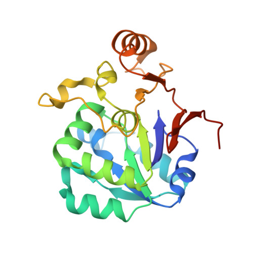
Catalytic triad heterogeneity in S51 peptidase family: Structural basis for functional variability.
Yadav, P., Goyal, V.D., Chandravanshi, K., Kumar, A., Gokhale, S.M., Jamdar, S.N., Makde, R.D.(2019) Proteins 87: 679-692
- PubMed: 30968972
- DOI: https://doi.org/10.1002/prot.25693
- Primary Citation of Related Structures:
6A4T, 6IRU - PubMed Abstract:
Peptidase E (PepE) is a nonclassical serine peptidase with a Ser-His-Glu catalytic triad. It is specific for dipeptides with an N-terminal aspartate residue (Asp-X dipeptidase activity). Its homolog from Listeria monocytogenes (PepElm) has a Ser-His-Asn "catalytic triad." Based on sequence alignment we predicted that the PepE homolog from Deinococcus radiodurans (PepEdr) would have a Ser-His-Asp "catalytic triad." We confirmed this by solving the crystal structure of PepEdr to 2.7 Å resolution. We show that PepElm and PepEdr lack the Asp-X dipeptidase activity. Our analyses suggest that absence of P1 pocket in the active site could be the main reason for this lack of typical activity. Sequence and structural data reveal that the PepE homologs can be divided into long and short PepEs based on presence or absence of a C-terminal tail which adopts a β-hairpin conformation in the canonical PepE from Salmonella enterica. A long PepE from Bacillus subtilis with Ser-His-Asp catalytic triad exhibits Asp-X dipeptidase activity. Whereas the three long PepEs enzymatically characterized till date have been found to possess the Asp-X dipeptidase activity, the three enzymatically characterized short PepEs lack this activity irrespective of the nature of their catalytic triads. This study illuminates the structural and functional heterogeneity in the S51 family and also provides structural basis for the functional variability among PepE homologs.
- High Pressure and Synchrotron Radiation Physics Division, Bhabha Atomic Research Centre, Mumbai, Maharashtra, India.
Organizational Affiliation:


