An isoform-selective inhibitor of tropomyosin receptor kinase A behaves as molecular glue.
Furuya, N., Momose, T., Katsuno, K., Fushimi, N., Muranaka, H., Handa, C., Sawa, M., Ozawa, T., Kinoshita, T.(2020) Bioorg Med Chem Lett 30: 126775-126775
- PubMed: 31699609 Search on PubMed
- DOI: https://doi.org/10.1016/j.bmcl.2019.126775
- Primary Citation Related Structures:
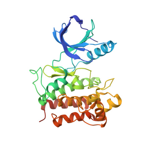
6IQN - PubMed Abstract:
The production of TrkA-selective inhibitors is considerably difficult because the kinase domains of TrkA and its isoforms TrkB/C have highly homologous amino acid sequences. Here we describe the structural basis for the acquisition of selectivity for a isoform-selective TrkA inhibitor, namely compound V1. The X-ray structure revealed that V1 acts as a molecular glue to stabilize the symmetrical dimer of the TrkA kinase domains. V1 binds to the ATP-binding site and simultaneously engages in the dimeric interface of TrkA. The region of the dimeric interface in TrkA is not conserved in TrkB/C; thus, dimer formation may be a novel mechanism for the production of selective TrkA inhibitors. The biochemical and biophysical assay results confirmed that V1 selectively inhibited TrkA and induced the dimer formation of TrkA, but not TrkB. The binding pocket at the TrkA dimer interface can be used for the production of new isoform-selective TrkA inhibitors.
- Kissei Pharmaceutical Inc., Azumino City, Nagano Pref., Japan; Graduate School of Science, Osaka Prefecture University, Osaka, Japan. Electronic address: noritaka_furuya@pharm.kissei.co.jp.
Organizational Affiliation: