6IQD | pdb_00006iqd

Crystal structure of Alcohol dehydrogenase from Geobacillus stearothermophilus


Experimental Data Snapshot

  • Method: X-RAY DIFFRACTION
  • Resolution: 2.84 Å
  • R-Value Free: 
    0.291 (Depositor), 0.291 (DCC) 
  • R-Value Work: 
    0.232 (Depositor), 0.235 (DCC) 
  • R-Value Observed: 
    0.235 (Depositor) 

Starting Model: experimental
View more details

wwPDB Validation 3D Report Full Report

Validation slider image for 6IQD

This is version 1.1 of the entry. See complete history

Literature

Characterization of the substrate scope of an alcohol dehydrogenase commonly used as methanol dehydrogenase.

Guo, X.Feng, Y.Wang, X.Liu, Y.Liu, W.Li, Q.Wang, J.Xue, S.Zhao, Z.K.

(2019) Bioorg Med Chem Lett 29: 1446-1449

  • DOI: https://doi.org/10.1016/j.bmcl.2019.04.025
  • Primary Citation Related Structures: 
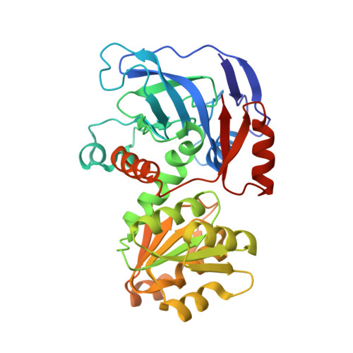
    6IQD

  • PubMed Abstract: 

    Many alcohol dehydrogenases (ADHs) catalyze oxidation of a broad scope of alcohols. When an NAD-dependent ADH oxidizes methanol, albeit at a poor rate, it may be treated as methanol dehydrogenase (MDH). One ADH from Geobacillus stearothermophilus DSM 2334 (GsADH) has been widely used as MDH, but its actual substrate scope remains less characterized. Here we purified recombinant GsADH from Escherichia coli and determined its crystal structure. We collected kinetics data of this enzyme towards a number of short chain alcohols, and found that isopropanol is by far the most favorable substrate. Moreover, molecular docking analysis suggested that substrate preference is mainly attributed to the conformer energy of the protein-substrate complex. Our data clarified the substrate scope of GsADH and provided structural insights, which may facilitate more efficient cofactor regeneration and rational metabolic engineering.


  • Organizational Affiliation
    • Dalian Institute of Chemical Physics, Chinese Academy of Sciences, Dalian 116023, China; University of Chinese Academy of Sciences, Beijing 100049, China.

Macromolecule Content 

  • Total Structure Weight: 291.07 kDa 
  • Atom Count: 20,152 
  • Modeled Residue Count: 2,688 
  • Deposited Residue Count: 2,712 
  • Unique protein chains: 1

Macromolecules

Find similar proteins by:|  3D Structure
Entity ID: 1
MoleculeChains  Sequence LengthOrganismDetailsImage
Alcohol dehydrogenase
A, B, C, D, E
A, B, C, D, E, F, G, H
339Geobacillus stearothermophilusMutation(s): 0 
Gene Names: adh
EC: 1.1.1.1
UniProt
Find proteins for P42327 (Geobacillus stearothermophilus)
Explore P42327 
Go to UniProtKB:  P42327
Entity Groups
Sequence Clusters30% Identity50% Identity70% Identity90% Identity95% Identity100% Identity
UniProt GroupP42327
Sequence Annotations
Expand
Reference Sequence

Small Molecules

Ligands 1 Unique
IDChains Name / Formula / InChI Key2D Diagram3D Interactions
ZN

Query on ZN


Download:Ideal Coordinates CCD File
I [auth A]
J [auth A]
K [auth B]
L [auth B]
M [auth C]
I [auth A],
J [auth A],
K [auth B],
L [auth B],
M [auth C],
N [auth C],
O [auth D],
P [auth D],
Q [auth E],
R [auth E],
S [auth F],
T [auth F],
U [auth G],
V [auth G],
W [auth H],
X [auth H]
ZINC ION
Zn
PTFCDOFLOPIGGS-UHFFFAOYSA-N

Experimental Data & Validation

Experimental Data

  • Method: X-RAY DIFFRACTION
  • Resolution: 2.84 Å
  • R-Value Free:  0.291 (Depositor), 0.291 (DCC) 
  • R-Value Work:  0.232 (Depositor), 0.235 (DCC) 
  • R-Value Observed: 0.235 (Depositor) 
Space Group: P 21 21 21
Unit Cell:
Length ( Å )Angle ( ˚ )
a = 71.442α = 90
b = 149.186β = 90
c = 269.277γ = 90
Software Package:
Software NamePurpose
PHENIXrefinement
HKL-3000data reduction
HKL-3000data scaling
PHASERphasing

Structure Validation

View Full Validation Report


Entry History 

Deposition Data

Revision History  (Full details and data files)

  • Version 1.0: 2019-06-05
    Type: Initial release
  • Version 1.1: 2023-11-22
    Changes: Data collection, Database references, Refinement description