6IQ6

Crystal structure of GAPDH


Experimental Data Snapshot

  • Method: X-RAY DIFFRACTION
  • Resolution: 2.29 Å
  • R-Value Free: 0.255 
  • R-Value Work: 0.215 
  • R-Value Observed: 0.217 

wwPDB Validation   3D Report Full Report


Ligand Structure Quality Assessment 


This is version 1.2 of the entry. See complete history


Literature

Structural Study of Monomethyl Fumarate-Bound Human GAPDH.

Park, J.B.Park, H.Son, J.Ha, S.J.Cho, H.S.

(2019) Mol Cells 42: 597-603

  • DOI: https://doi.org/10.14348/molcells.2019.0114
  • Primary Citation of Related Structures:  
    6IQ6

  • PubMed Abstract: 

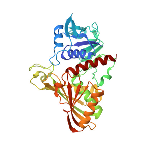
    Glyceraldehyde-3-phosphate dehydrogenase (GAPDH) is a core enzyme of the aerobic glycolytic pathway with versatile functions and is associated with cancer development. Recently, Kornberg et al . published the detailed correlation between GAPDH and di - or monomethyl fumarate (DMF or MMF), which are well-known GAPDH antagonists in the immune system. As an extension, herein, we report the crystal structure of MMF-bound human GAPDH at 2.29 Å. The MMF molecule is covalently linked to the catalytic Cys152 of human GAPDH, and inhibits the catalytic activity of the residue and dramatically reduces the enzymatic activity of GAPDH. Structural comparisons between NAD + bound GAPDH and MMF-bound GAPDH revealed that the covalently linked MMF can block the binding of the NAD + cosubstrate due to steric hindrance of the nicotinamide portion of the NAD + molecule, illuminating the specific mechanism by which MMF inhibits GAPDH. Our data provide insights into GAPDH antagonist development for GAPDH-mediated disease treatment.


  • Organizational Affiliation

    Department of Systems Biology, College of Life Science and Biotechnology, Yonsei University, Seoul 03722, Korea.


Macromolecules
Find similar proteins by:  (by identity cutoff)  |  3D Structure
Entity ID: 1
MoleculeChains Sequence LengthOrganismDetailsImage
Glyceraldehyde-3-phosphate dehydrogenase
A, B, C, D, E
A, B, C, D, E, F, G, H
335Homo sapiensMutation(s): 0 
Gene Names: GAPDHGAPDCDABP0047OK/SW-cl.12
EC: 1.2.1.12 (PDB Primary Data), 2.6.99 (PDB Primary Data)
UniProt & NIH Common Fund Data Resources
Find proteins for P04406 (Homo sapiens)
Explore P04406 
Go to UniProtKB:  P04406
PHAROS:  P04406
GTEx:  ENSG00000111640 
Entity Groups  
Sequence Clusters30% Identity50% Identity70% Identity90% Identity95% Identity100% Identity
UniProt GroupP04406
Sequence Annotations
Expand
  • Reference Sequence
Experimental Data & Validation

Experimental Data

  • Method: X-RAY DIFFRACTION
  • Resolution: 2.29 Å
  • R-Value Free: 0.255 
  • R-Value Work: 0.215 
  • R-Value Observed: 0.217 
  • Space Group: P 1 21 1
Unit Cell:
Length ( Å )Angle ( ˚ )
a = 71.63α = 90
b = 108.49β = 96.37
c = 175.81γ = 90
Software Package:
Software NamePurpose
REFMACrefinement
HKL-2000data reduction
HKL-2000data scaling
MOLREPphasing

Structure Validation

View Full Validation Report


Ligand Structure Quality Assessment 


Entry History 

Deposition Data

Revision History  (Full details and data files)

  • Version 1.0: 2019-08-28
    Type: Initial release
  • Version 1.1: 2019-09-11
    Changes: Data collection, Database references
  • Version 1.2: 2023-11-22
    Changes: Data collection, Database references, Refinement description