Antimicrobial silver targets glyceraldehyde-3-phosphate dehydrogenase in glycolysis ofE. coli.
Wang, H., Wang, M., Yang, X., Xu, X., Hao, Q., Yan, A., Hu, M., Lobinski, R., Li, H., Sun, H.(2019) Chem Sci 10: 7193-7199
- PubMed: 31588287 Search on PubMedSearch on PubMed Central
- DOI: https://doi.org/10.1039/c9sc02032b
- Primary Citation Related Structures:
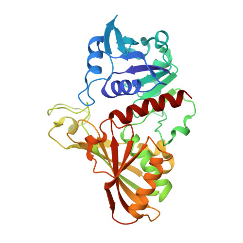
6IO4, 6IO6, 6IOJ - PubMed Abstract:
Silver has long been used as an antibacterial agent, yet its molecular targets remain largely unknown. Using a custom-designed coupling of gel electrophoresis with inductively coupled plasma mass spectrometry (GE-ICP-MS), we identified six silver-binding proteins in E. coli . The majority of the identified proteins are associated with the central carbon metabolism of E. coli . Among them, we unveil that GAPDH, an essential enzyme in glycolysis, serves as a vital target of Ag + in E. coli for the first time. We demonstrate that silver inhibits the enzymatic function of GAPDH through targeting Cys149 in its catalytic site. The X-ray structure reveals that Ag + coordinates to Cys149 and His176 with a quasi-linear geometry (S-Ag-N angle of 157°). And unexpectedly, two Ag + ions coordinate to Cys288 in the non-catalytic site with weak argentophilic interaction (Ag···Ag distance of 2.9 Å). This is the first report on antimicrobial Ag + targeting a key enzyme in the glycolytic pathway of E. coli . The findings expand our knowledge on the mode of action and bio-coordination chemistry of silver, particularly silver-targeting residues in proteins at the atomic level.
- Department of Chemistry , The University of Hong Kong , Hong Kong , P. R. China . Email: hsun@hku.hk.
Organizational Affiliation: