


Crown Ethers as Transthyretin Amyloidogenesis Inhibitor
Yokoyama, T., Kosaka, Y., Matsumoto, K., Kitakami, R., Nabeshima, Y., Mizuguchi, M.To be published.
Experimental Data Snapshot
Entity ID: 1 | |||||
|---|---|---|---|---|---|
| Molecule | Chains | Sequence Length | Organism | Details | Image |
| Histone-lysine N-methyltransferase, H3 lysine-79 specific | 336 | Homo sapiens | Mutation(s): 0 Gene Names: DOT1L, KIAA1814, KMT4 EC: 2.1.1.43 (PDB Primary Data), 2.1.1.360 (UniProt) | 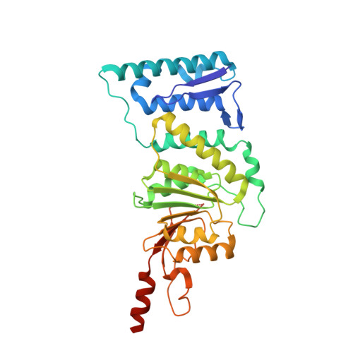 | |
UniProt & NIH Common Fund Data Resources | |||||
PHAROS: Q8TEK3 GTEx: ENSG00000104885 | |||||
Entity Groups | |||||
| Sequence Clusters | 30% Identity50% Identity70% Identity90% Identity95% Identity100% Identity | ||||
| UniProt Group | Q8TEK3 | ||||
Sequence AnnotationsExpand | |||||
Reference Sequence | |||||
| Ligands 3 Unique | |||||
|---|---|---|---|---|---|
| ID | Chains | Name / Formula / InChI Key | 2D Diagram | 3D Interactions | |
| SAH Download:Ideal Coordinates CCD File | C [auth A] | S-ADENOSYL-L-HOMOCYSTEINE C14 H20 N6 O5 S ZJUKTBDSGOFHSH-WFMPWKQPSA-N |  | ||
| O4B Download:Ideal Coordinates CCD File | B [auth A] | 1,4,7,10,13,16-HEXAOXACYCLOOCTADECANE C12 H24 O6 XEZNGIUYQVAUSS-UHFFFAOYSA-N |  | ||
| SO4 Download:Ideal Coordinates CCD File | D [auth A], E [auth A], F [auth A], G [auth A], H [auth A] | SULFATE ION O4 S QAOWNCQODCNURD-UHFFFAOYSA-L |  | ||
| Length ( Å ) | Angle ( ˚ ) |
|---|---|
| a = 145.092 | α = 90 |
| b = 145.092 | β = 90 |
| c = 56.478 | γ = 120 |
| Software Name | Purpose |
|---|---|
| PHENIX | refinement |
| XDS | data reduction |
| XSCALE | data scaling |
| MOLREP | phasing |


