Phagocytosis is mediated by two-dimensional assemblies of the F-BAR protein GAS7.
Hanawa-Suetsugu, K., Itoh, Y., Ab Fatah, M., Nishimura, T., Takemura, K., Takeshita, K., Kubota, S., Miyazaki, N., Wan Mohamad Noor, W.N.I., Inaba, T., Nguyen, N.T.H., Hamada-Nakahara, S., Oono-Yakura, K., Tachikawa, M., Iwasaki, K., Kohda, D., Yamamoto, M., Kitao, A., Shimada, A., Suetsugu, S.(2019) Nat Commun 10: 4763-4763
- PubMed: 31628328 Search on PubMedSearch on PubMed Central
- DOI: https://doi.org/10.1038/s41467-019-12738-w
- Primary Citation Related Structures:
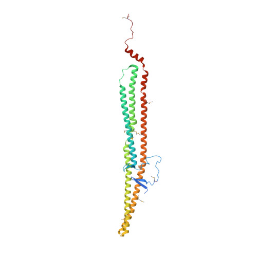
6IKN, 6IKO - PubMed Abstract:
Phagocytosis is a cellular process for internalization of micron-sized large particles including pathogens. The Bin-Amphiphysin-Rvs167 (BAR) domain proteins, including the FCH-BAR (F-BAR) domain proteins, impose specific morphologies on lipid membranes. Most BAR domain proteins are thought to form membrane invaginations or protrusions by assembling into helical submicron-diameter filaments, such as on clathrin-coated pits, caveolae, and filopodia. However, the mechanism by which BAR domain proteins assemble into micron-scale phagocytic cups was unclear. Here, we show that the two-dimensional sheet-like assembly of Growth Arrest-Specific 7 (GAS7) plays a critical role in phagocytic cup formation in macrophages. GAS7 has the F-BAR domain that possesses unique hydrophilic loops for two-dimensional sheet formation on flat membranes. Super-resolution microscopy reveals the similar assemblies of GAS7 on phagocytic cups and liposomes. The mutations of the loops abolishes both the membrane localization of GAS7 and phagocytosis. Thus, the sheet-like assembly of GAS7 plays a significant role in phagocytosis.
- Nara Institute of Science and Technology, Ikoma, 630-0192, Japan.
Organizational Affiliation: