6IEX | pdb_00006iex

Crystal structure of HLA-B*4001 in complex with SARS-CoV derived peptide N216-225 GETALALLLL


Experimental Data Snapshot

  • Method: X-RAY DIFFRACTION
  • Resolution: 2.31 Å
  • R-Value Free: 
    0.261 (Depositor), 0.254 (DCC) 
  • R-Value Work: 
    0.211 (Depositor), 0.208 (DCC) 
  • R-Value Observed: 
    0.213 (Depositor) 

wwPDB Validation 3D Report Full Report

Validation slider image for 6IEX

This is version 1.2 of the entry. See complete history

Literature

Salt bridge-forming residues positioned over viral peptides presented by MHC class I impacts T-cell recognition in a binding-dependent manner.

Ji, W.Niu, L.Peng, W.Zhang, Y.Cheng, H.Gao, F.Shi, Y.Qi, J.Gao, G.F.Liu, W.J.

(2019) Mol Immunol 112: 274-282

  • DOI: https://doi.org/10.1016/j.molimm.2019.06.005
  • Primary Citation Related Structures: 
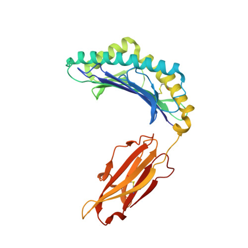
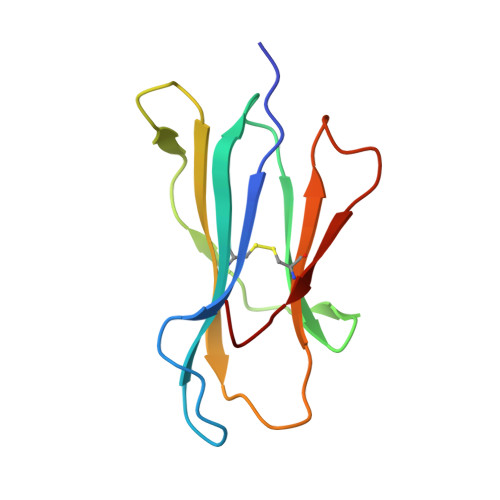
    6IEX

  • PubMed Abstract: 

    The viral peptides presentation by major histocompatibility complex class I (MHC I) molecules play a pivotal role in T-cell recognition and the subsequent virus clearance. This process is delicately adjusted by the variant residues of MHC I, especially the residues in the peptide binding groove (PBG). In a series of MHC I molecules, a salt bridge is formed above the N-terminus of the peptides. However, the potential impact of the salt bridge on peptide binding and T-cell receptor (TCR) recognition of MHC I, as well as the corresponding molecular basis, are still largely unknown. Herein, we determined the structures of HLA-B*4001 and H-2K d in which two different types of salt bridges (Arg62-Glu163 or Arg66-Glu163) across the PBG were observed. Although the two salt bridges led to different conformation shifts of both the MHC I α helix and the peptides, binding of the peptides with the salt bridge residues was relatively conserved. Furthermore, through a series of in vitro and in vivo investigations, we found that MHC I mutations that disrupt the salt bridge alleviate peptide binding and can weaken the TCR recognition of MHC I-peptide complexes. Our study may provide key references for understanding MHC I-restricted peptide recognition by T-cells.


  • Organizational Affiliation
    • NHC Key Laboratory of Medical Virology and Viral Diseases, National Institute for Viral Disease Control and Prevention, Chinese Center for Disease Control and Prevention, Beijing 100052, China.

Macromolecule Content 

  • Total Structure Weight: 44.69 kDa 
  • Atom Count: 3,255 
  • Modeled Residue Count: 383 
  • Deposited Residue Count: 384 
  • Unique protein chains: 3

Macromolecules

Find similar proteins by:|  3D Structure
Entity ID: 1
MoleculeChains  Sequence LengthOrganismDetailsImage
MHC class I antigen274Homo sapiensMutation(s): 0 
Gene Names: HLA-B
UniProt
Find proteins for A0A499S0B6 (Homo sapiens)
Explore A0A499S0B6 
Go to UniProtKB:  A0A499S0B6
Entity Groups
Sequence Clusters30% Identity50% Identity70% Identity90% Identity95% Identity100% Identity
UniProt GroupA0A499S0B6
Sequence Annotations
Expand
Reference Sequence
Find similar proteins by:|  3D Structure
Entity ID: 2
MoleculeChains  Sequence LengthOrganismDetailsImage
Beta-2-microglobulin100Homo sapiensMutation(s): 0 
Gene Names: B2M
UniProt & NIH Common Fund Data Resources
Find proteins for P61769 (Homo sapiens)
Explore P61769 
Go to UniProtKB:  P61769
PHAROS:  P61769
GTEx:  ENSG00000166710 
Entity Groups
Sequence Clusters30% Identity50% Identity70% Identity90% Identity95% Identity100% Identity
UniProt GroupP61769
Sequence Annotations
Expand
Reference Sequence
Find similar proteins by:  Sequence   |   3D Structure  
Entity ID: 3
MoleculeChains  Sequence LengthOrganismDetailsImage
GLY-GLU-THR-ALA-LEU-ALA-LEU-LEU-LEU-LEU10Severe acute respiratory syndrome-related coronavirusMutation(s): 0 
UniProt
Find proteins for P59595 (Severe acute respiratory syndrome coronavirus)
Explore P59595 
Go to UniProtKB:  P59595
Entity Groups
Sequence Clusters30% Identity50% Identity70% Identity90% Identity95% Identity100% Identity
UniProt GroupP59595
Sequence Annotations
Expand
Reference Sequence

Experimental Data & Validation

Experimental Data

  • Method: X-RAY DIFFRACTION
  • Resolution: 2.31 Å
  • R-Value Free:  0.261 (Depositor), 0.254 (DCC) 
  • R-Value Work:  0.211 (Depositor), 0.208 (DCC) 
  • R-Value Observed: 0.213 (Depositor) 
Space Group: P 21 21 21
Unit Cell:
Length ( Å )Angle ( ˚ )
a = 52.482α = 90
b = 69.274β = 90
c = 120.936γ = 90
Software Package:
Software NamePurpose
PHENIXrefinement
XDSdata reduction
XDSdata scaling
CNSphasing

Structure Validation

View Full Validation Report


Entry History 

Deposition Data

Revision History  (Full details and data files)

  • Version 1.0: 2019-09-18
    Type: Initial release
  • Version 1.1: 2021-03-31
    Changes: Database references
  • Version 1.2: 2024-10-16
    Changes: Advisory, Data collection, Database references, Structure summary