6IED

Crystal structure of heme A synthase from Bacillus subtilis


Experimental Data Snapshot

  • Method: X-RAY DIFFRACTION
  • Resolution: 3.00 Å
  • R-Value Free: 0.245 
  • R-Value Work: 0.233 

wwPDB Validation   3D Report Full Report


Ligand Structure Quality Assessment 


This is version 1.2 of the entry. See complete history


Literature

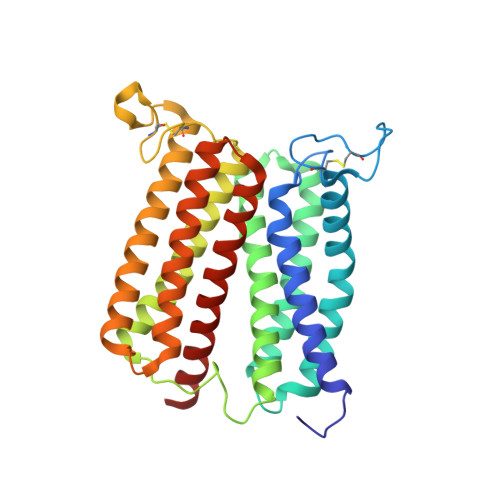
Crystal structure of heme A synthase fromBacillus subtilis.

Niwa, S.Takeda, K.Kosugi, M.Tsutsumi, E.Mogi, T.Miki, K.

(2018) Proc Natl Acad Sci U S A 115: 11953-11957

  • DOI: https://doi.org/10.1073/pnas.1813346115
  • Primary Citation of Related Structures:  
    6A2J, 6IED

  • PubMed Abstract: 

    Heme A is an essential cofactor for respiratory terminal oxidases and vital for respiration in aerobic organisms. The final step of heme A biosynthesis is formylation of the C-8 methyl group of heme molecule by heme A synthase (HAS). HAS is a heme-containing integral membrane protein, and its structure and reaction mechanisms have remained unknown. Thus, little is known about HAS despite of its importance. Here we report the crystal structure of HAS from Bacillus subtilis at 2.2-Å resolution. The N- and C-terminal halves of HAS consist of four-helix bundles and they align in a pseudo twofold symmetry manner. Each bundle contains a pair of histidine residues and forms a heme-binding domain. The C-half domain binds a cofactor-heme molecule, while the N-half domain is vacant. Many water molecules are found in the transmembrane region and around the substrate-binding site, and some of them interact with the main chain of transmembrane helix. Comparison of these two domain structures enables us to construct a substrate-heme binding state structure. This structure implies that a completely conserved glutamate, Glu57 in B. subtilis , is the catalytic residue for the formylation reaction. These results provide valuable suggestions of the substrate-heme binding mechanism. Our results present significant insight into the heme A biosynthesis.


  • Organizational Affiliation

    Department of Chemistry, Graduate School of Science, Kyoto University, 606-8502 Kyoto, Japan.


Macromolecules
Find similar proteins by:  (by identity cutoff)  |  3D Structure
Entity ID: 1
MoleculeChains Sequence LengthOrganismDetailsImage
Heme A synthase309Bacillus subtilis subsp. subtilis str. 168Mutation(s): 0 
Gene Names: ctaABSU14870
EC: 1.3
Membrane Entity: Yes 
UniProt
Find proteins for P12946 (Bacillus subtilis (strain 168))
Explore P12946 
Go to UniProtKB:  P12946
Entity Groups  
Sequence Clusters30% Identity50% Identity70% Identity90% Identity95% Identity100% Identity
UniProt GroupP12946
Sequence Annotations
Expand
  • Reference Sequence
Small Molecules
Experimental Data & Validation

Experimental Data

  • Method: X-RAY DIFFRACTION
  • Resolution: 3.00 Å
  • R-Value Free: 0.245 
  • R-Value Work: 0.233 
  • Space Group: H 3
Unit Cell:
Length ( Å )Angle ( ˚ )
a = 90.528α = 90
b = 90.528β = 90
c = 147.23γ = 120
Software Package:
Software NamePurpose
DENZOdata reduction
SCALEPACKdata scaling
CNSphasing
CNSrefinement

Structure Validation

View Full Validation Report


Ligand Structure Quality Assessment 


Entry History & Funding Information

Deposition Data


Funding OrganizationLocationGrant Number
Ministry of Education, Culture, Sports, Science and Technology (Japan)Japan--
Japan Society for the Promotion of ScienceJapan15J02413

Revision History  (Full details and data files)

  • Version 1.0: 2018-11-21
    Type: Initial release
  • Version 1.1: 2018-12-12
    Changes: Data collection, Database references
  • Version 1.2: 2023-11-22
    Changes: Data collection, Database references, Derived calculations, Refinement description