


Crystal Structure of the first bromodomain of BRD4 in complex with RT56
Picaud, S., Traquete, R., Bernardes, G.J.L., Newman, J., Arrowsmith, C.H., Edwards, A.M., Bountra, C., Filippakopoulos, P., Structural Genomics Consortium (SGC)To be published.
Experimental Data Snapshot
Starting Model: experimental
View more details
Entity ID: 1 | |||||
|---|---|---|---|---|---|
| Molecule | Chains | Sequence Length | Organism | Details | Image |
| Bromodomain-containing protein 4 | 127 | Homo sapiens | Mutation(s): 0 Gene Names: BRD4, HUNK1 | 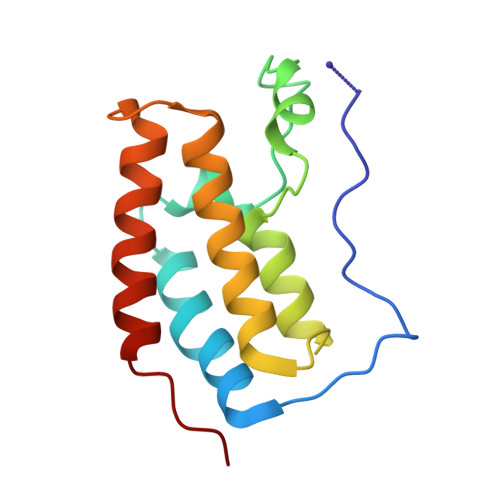 | |
UniProt & NIH Common Fund Data Resources | |||||
PHAROS: O60885 GTEx: ENSG00000141867 | |||||
Entity Groups | |||||
| Sequence Clusters | 30% Identity50% Identity70% Identity90% Identity95% Identity100% Identity | ||||
| UniProt Group | O60885 | ||||
Sequence AnnotationsExpand | |||||
Reference Sequence | |||||
| Ligands 2 Unique | |||||
|---|---|---|---|---|---|
| ID | Chains | Name / Formula / InChI Key | 2D Diagram | 3D Interactions | |
| H7E Download:Ideal Coordinates CCD File | D [auth A] | [1-[4-[2-[(4~{S})-6-(4-chlorophenyl)-8-methoxy-1-methyl-4~{H}-[1,2,4]triazolo[4,3-a][1,4]benzodiazepin-4-yl]ethanoylamino]phenyl]piperidin-4-yl]-trimethyl-azanium C34 H39 Cl N7 O2 WDWNSWKCDGLBCQ-PMERELPUSA-O |  | ||
| EDO Download:Ideal Coordinates CCD File | B [auth A], C [auth A] | 1,2-ETHANEDIOL C2 H6 O2 LYCAIKOWRPUZTN-UHFFFAOYSA-N |  | ||
| Length ( Å ) | Angle ( ˚ ) |
|---|---|
| a = 37.738 | α = 90 |
| b = 44.739 | β = 90 |
| c = 79.563 | γ = 90 |
| Software Name | Purpose |
|---|---|
| SCALA | data scaling |
| PHASER | phasing |
| REFMAC | refinement |
| PDB_EXTRACT | data extraction |
| XDS | data reduction |
| Funding Organization | Location | Grant Number |
|---|---|---|
| Wellcome Trust | United Kingdom | 095751/Z/11/Z |


