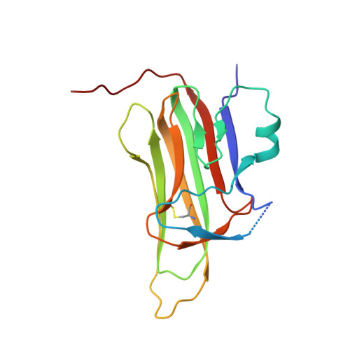
6I50

Structure of Eiger TNF from S. frugiperda


Experimental Data Snapshot

  • Method: X-RAY DIFFRACTION
  • Resolution: 1.69 Å
  • R-Value Free: 0.182 
  • R-Value Work: 0.155 
  • R-Value Observed: 0.157 

Starting Model: experimental
View more details

wwPDB Validation   3D Report Full Report


This is version 1.3 of the entry. See complete history


Literature

High-resolution crystal structure of arthropod Eiger TNF suggests a mode of receptor engagement and altered surface charge within endosomes.

Bertinelli, M.Paesen, G.C.Grimes, J.M.Renner, M.

(2019) Commun Biol 2: 293-293

  • DOI: https://doi.org/10.1038/s42003-019-0541-0
  • Primary Citation of Related Structures:  
    6I50

  • PubMed Abstract: 

    The tumour necrosis factor alpha (TNFα) superfamily of proteins are critical in numerous biological processes, such as in development and immunity. Eiger is the sole TNFα member described in arthropods such as in the important model organism Drosophila . To date there are no structural data on any Eiger protein. Here we present the structure of the TNF domain of Eiger from the fall armyworm Spodoptera frugiperda (SfEiger) to 1.7 Å from a serendipitously obtained crystal without prior knowledge of the protein sequence. Our structure confirms that canonical trimerization is conserved from ancestral TNFs and points towards a mode of receptor engagement. Furthermore, we observe numerous surface histidines on SfEiger, potentially acting as pH switches following internalization into endosomes. Our data contributes to the genome annotation of S. frugiperda , a voracious agricultural pest, and can serve as a basis for future structure-function investigations of the TNF system in related arthropods such as Drosophila .


  • Organizational Affiliation

    1Division of Structural Biology, Wellcome Centre for Human Genetics, University of Oxford, 10 Roosevelt Drive, Oxford, OX3 7BN UK.


Macromolecules
Find similar proteins by:  (by identity cutoff)  |  3D Structure
Entity ID: 1
MoleculeChains Sequence LengthOrganismDetailsImage
SFRICE_029225170Spodoptera frugiperdaMutation(s): 0 
UniProt
Find proteins for A0A2H1VTZ0 (Spodoptera frugiperda)
Explore A0A2H1VTZ0 
Go to UniProtKB:  A0A2H1VTZ0
Entity Groups  
Sequence Clusters30% Identity50% Identity70% Identity90% Identity95% Identity100% Identity
UniProt GroupA0A2H1VTZ0
Sequence Annotations
Expand
  • Reference Sequence
Experimental Data & Validation

Experimental Data

  • Method: X-RAY DIFFRACTION
  • Resolution: 1.69 Å
  • R-Value Free: 0.182 
  • R-Value Work: 0.155 
  • R-Value Observed: 0.157 
  • Space Group: H 3
Unit Cell:
Length ( Å )Angle ( ˚ )
a = 55.222α = 90
b = 55.222β = 90
c = 142.096γ = 120
Software Package:
Software NamePurpose
PHENIXrefinement
DIALSdata reduction
DIALSdata scaling
PHASERphasing

Structure Validation

View Full Validation Report


Entry History & Funding Information

Deposition Data


Funding OrganizationLocationGrant Number
Wellcome TrustUnited Kingdom204703/Z/16/Z

Revision History  (Full details and data files)

  • Version 1.0: 2019-08-14
    Type: Initial release
  • Version 1.1: 2019-08-28
    Changes: Data collection, Database references
  • Version 1.2: 2019-11-20
    Changes: Database references
  • Version 1.3: 2024-01-24
    Changes: Data collection, Database references, Refinement description