6I4S

Crystal structure of the disease-causing R447G mutant of the human dihydrolipoamide dehydrogenase


Experimental Data Snapshot

  • Method: X-RAY DIFFRACTION
  • Resolution: 1.75 Å
  • R-Value Free: 0.194 
  • R-Value Work: 0.178 
  • R-Value Observed: 0.179 

wwPDB Validation   3D Report Full Report


Ligand Structure Quality Assessment 


This is version 1.2 of the entry. See complete history


Literature

Underlying molecular alterations in human dihydrolipoamide dehydrogenase deficiency revealed by structural analyses of disease-causing enzyme variants.

Szabo, E.Wilk, P.Nagy, B.Zambo, Z.Bui, D.Weichsel, A.Arjunan, P.Torocsik, B.Hubert, A.Furey, W.Montfort, W.R.Jordan, F.Weiss, M.S.Adam-Vizi, V.Ambrus, A.

(2019) Hum Mol Genet 28: 3339-3354

  • DOI: https://doi.org/10.1093/hmg/ddz177
  • Primary Citation of Related Structures:  
    6I4P, 6I4Q, 6I4R, 6I4S, 6I4T, 6I4U, 6I4Z

  • PubMed Abstract: 

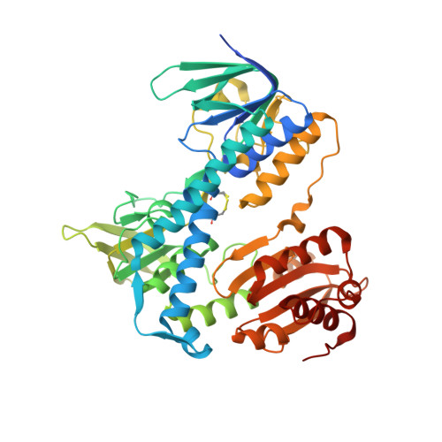
    Human dihydrolipoamide dehydrogenase (hLADH, hE3) deficiency (OMIM# 246900) is an often prematurely lethal genetic disease usually caused by inactive or partially inactive hE3 variants. Here we report the crystal structure of wild-type hE3 at an unprecedented high resolution of 1.75 Å and the structures of six disease-causing hE3 variants at resolutions ranging from 1.44 to 2.34 Å. P453L proved to be the most deleterious substitution in structure as aberrations extensively compromised the active site. The most prevalent G194C-hE3 variant primarily exhibited structural alterations close to the substitution site, whereas the nearby cofactor-binding residues were left unperturbed. The G426E substitution mainly interfered with the local charge distribution introducing dynamics to the substitution site in the dimer interface; G194C and G426E both led to minor structural changes. The R460G, R447G and I445M substitutions all perturbed a solvent accessible channel, the so-called H+/H2O channel, leading to the active site. Molecular pathomechanisms of enhanced reactive oxygen species (ROS) generation and impaired binding to multienzyme complexes were also addressed according to the structural data for the relevant mutations. In summary, we present here for the first time a comprehensive study that links three-dimensional structures of disease-causing hE3 variants to residual hLADH activities, altered capacities for ROS generation, compromised affinities for multienzyme complexes and eventually clinical symptoms. Our results may serve as useful starting points for future therapeutic intervention approaches.


  • Organizational Affiliation

    Department of Medical Biochemistry, MTA-SE Laboratory for Neurobiochemistry, Semmelweis University, Budapest, 1094, Hungary.


Macromolecules
Find similar proteins by:  (by identity cutoff)  |  3D Structure
Entity ID: 1
MoleculeChains Sequence LengthOrganismDetailsImage
Dihydrolipoyl dehydrogenase, mitochondrial
A, B
496Homo sapiensMutation(s): 1 
Gene Names: DLDGCSLLADPHE3
EC: 1.8.1.4
UniProt & NIH Common Fund Data Resources
Find proteins for P09622 (Homo sapiens)
Explore P09622 
Go to UniProtKB:  P09622
PHAROS:  P09622
GTEx:  ENSG00000091140 
Entity Groups  
Sequence Clusters30% Identity50% Identity70% Identity90% Identity95% Identity100% Identity
UniProt GroupP09622
Sequence Annotations
Expand
  • Reference Sequence
Small Molecules
Ligands 2 Unique
IDChains Name / Formula / InChI Key2D Diagram3D Interactions
FAD
Query on FAD

Download Ideal Coordinates CCD File 
C [auth A],
I [auth B]
FLAVIN-ADENINE DINUCLEOTIDE
C27 H33 N9 O15 P2
VWWQXMAJTJZDQX-UYBVJOGSSA-N
SO4
Query on SO4

Download Ideal Coordinates CCD File 
D [auth A]
E [auth A]
F [auth A]
G [auth A]
H [auth A]
D [auth A],
E [auth A],
F [auth A],
G [auth A],
H [auth A],
J [auth B],
K [auth B],
L [auth B],
M [auth B],
N [auth B]
SULFATE ION
O4 S
QAOWNCQODCNURD-UHFFFAOYSA-L
Experimental Data & Validation

Experimental Data

  • Method: X-RAY DIFFRACTION
  • Resolution: 1.75 Å
  • R-Value Free: 0.194 
  • R-Value Work: 0.178 
  • R-Value Observed: 0.179 
  • Space Group: P 21 21 2
Unit Cell:
Length ( Å )Angle ( ˚ )
a = 117.273α = 90
b = 169.043β = 90
c = 60.845γ = 90
Software Package:
Software NamePurpose
MxCuBEdata collection
XDSdata reduction
XDSdata scaling
MOLREPphasing
PHENIXrefinement
PDB_EXTRACTdata extraction

Structure Validation

View Full Validation Report


Ligand Structure Quality Assessment 


Entry History & Funding Information

Deposition Data


Funding OrganizationLocationGrant Number
Hungarian Academy of SciencesHungary02001 [to A.-V.V.]
HungaryHungarian Scientific Research Fund, grant#112230 [to A.-V.V.]
HungaryHungarian Brain Research Program, grant#KTIA_13_NAP_III/6 and 2017-1.2.1-NKP-2017-00002 [to A.-V.V.]
Hungarian Academy of SciencesHungaryBolyai Fellowship [to A.A.]
European Molecular Biology OrganizationHungaryShort-term Fellowship [to A.A.]
HungarySemmelweis University, Young Investigator Research Grant [to A.A.]
HungaryGedeon Richter PIc., Young Investigator Research Grant [to A.A.]
United StatesFulbright Commission, Fulbright Fellowship [to A.A.]
HungaryEuropean Union and Government of Hungary, grant#EFOP-3.6.3-VEKOP-16-2017-00009 [to S.E.]
GermanyEuropean Union CALIPSOplus, grant#16204087-ST [to S.E. and A.A.]

Revision History  (Full details and data files)

  • Version 1.0: 2019-11-20
    Type: Initial release
  • Version 1.1: 2019-12-11
    Changes: Database references
  • Version 1.2: 2024-01-24
    Changes: Data collection, Database references, Refinement description