Equatorial Active Site Compaction and Electrostatic Reorganization in Catechol-O-methyltransferase.
Czarnota, S., Johannissen, L.O., Baxter, N.J., Rummel, F., Wilson, A.L., Cliff, M.J., Levy, C.W., Scrutton, N.S., Waltho, J.P., Hay, S.(2019) ACS Catal 9: 4394-4401
- PubMed: 31080692 Search on PubMedSearch on PubMed Central
- DOI: https://doi.org/10.1021/acscatal.9b00174
- Primary Citation Related Structures:
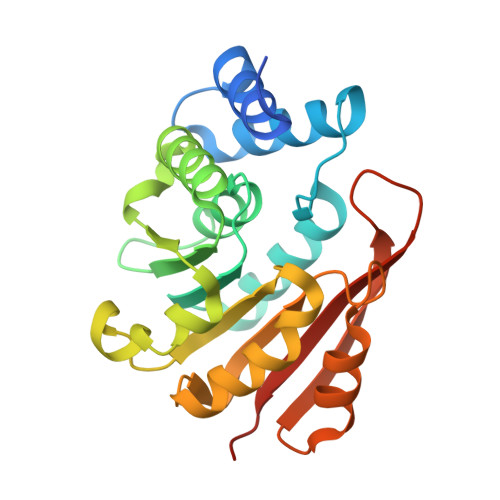
6I3C, 6I3D - PubMed Abstract:
Catechol- O -methyltransferase (COMT) is a model S-adenosyl-l-methionine (SAM) dependent methyl transferase, which catalyzes the methylation of catecholamine neurotransmitters such as dopamine in the primary pathway of neurotransmitter deactivation in animals. Despite extensive study, there is no consensus view of the physical basis of catalysis in COMT. Further progress requires experimental data that directly probes active site geometry, protein dynamics and electrostatics, ideally in a range of positions along the reaction coordinate. Here we establish that sinefungin, a fungal-derived inhibitor of SAM-dependent enzymes that possess transition state-like charge on the transferring group, can be used as a transition state analog of COMT when combined with a catechol. X-ray crystal structures and NMR backbone assignments of the ternary complexes of the soluble form of human COMT containing dinitrocatechol, Mg 2+ and SAM or sinefungin were determined. Comparison and further analysis with the aid of density functional theory calculations and molecular dynamics simulations provides evidence for active site "compaction", which is driven by electrostatic stabilization between the transferring methyl group and "equatorial" active site residues that are orthogonal to the donor-acceptor (pseudo reaction) coordinate. We propose that upon catecholamine binding and subsequent proton transfer to Lys 144, the enzyme becomes geometrically preorganized, with little further movement along the donor-acceptor coordinate required for methyl transfer. Catalysis is then largely facilitated through stabilization of the developing charge on the transferring methyl group via "equatorial" H-bonding and electrostatic interactions orthogonal to the donor-acceptor coordinate.
- Manchester Institute of Biotechnology, The University of Manchester, 131 Princess Street, Manchester, M1 7DN, United Kingdom.
Organizational Affiliation: