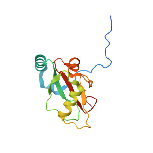
6HYO

Structure of ULK1 LIR motif bound to GABARAP


Experimental Data Snapshot

  • Method: X-RAY DIFFRACTION
  • Resolution: 1.07 Å
  • R-Value Free: 0.161 
  • R-Value Work: 0.142 
  • R-Value Observed: 0.142 

wwPDB Validation   3D Report Full Report


This is version 1.2 of the entry. See complete history


Literature

Molecular determinants regulating selective binding of autophagy adapters and receptors to ATG8 proteins.

Wirth, M.Zhang, W.Razi, M.Nyoni, L.Joshi, D.O'Reilly, N.Johansen, T.Tooze, S.A.Mouilleron, S.

(2019) Nat Commun 10: 2055-2055

  • DOI: https://doi.org/10.1038/s41467-019-10059-6
  • Primary Citation of Related Structures:  
    6HYL, 6HYM, 6HYN, 6HYO

  • PubMed Abstract: 

    Autophagy is an essential recycling and quality control pathway. Mammalian ATG8 proteins drive autophagosome formation and selective removal of protein aggregates and organelles by recruiting autophagy receptors and adaptors that contain a LC3-interacting region (LIR) motif. LIR motifs can be highly selective for ATG8 subfamily proteins (LC3s/GABARAPs), however the molecular determinants regulating these selective interactions remain elusive. Here we show that residues within the core LIR motif and adjacent C-terminal region as well as ATG8 subfamily-specific residues in the LIR docking site are critical for binding of receptors and adaptors to GABARAPs. Moreover, rendering GABARAP more LC3B-like impairs autophagy receptor degradation. Modulating LIR binding specificity of the centriolar satellite protein PCM1, implicated in autophagy and centrosomal function, alters its dynamics in cells. Our data provides new mechanistic insight into how selective binding of LIR motifs to GABARAPs is achieved, and elucidate the overlapping and distinct functions of ATG8 subfamily proteins.


  • Organizational Affiliation

    Molecular Cell Biology of Autophagy, The Francis Crick Institute, 1 Midland Road, London, NW1 1AT, UK. martina.wirth@crick.ac.uk.


Macromolecules
Find similar proteins by:  (by identity cutoff)  |  3D Structure
Entity ID: 1
MoleculeChains Sequence LengthOrganismDetailsImage
Serine/threonine-protein kinase ULK1,Gamma-aminobutyric acid receptor-associated protein137Homo sapiensMutation(s): 0 
Gene Names: ULK1KIAA0722GABARAPFLC3BHT004
EC: 2.7.11.1
UniProt & NIH Common Fund Data Resources
Find proteins for O95166 (Homo sapiens)
Explore O95166 
Go to UniProtKB:  O95166
PHAROS:  O95166
GTEx:  ENSG00000170296 
Find proteins for O75385 (Homo sapiens)
Explore O75385 
Go to UniProtKB:  O75385
PHAROS:  O75385
GTEx:  ENSG00000177169 
Entity Groups  
Sequence Clusters30% Identity50% Identity70% Identity90% Identity95% Identity100% Identity
UniProt GroupsO95166O75385
Sequence Annotations
Expand
  • Reference Sequence
Small Molecules
Ligands 3 Unique
IDChains Name / Formula / InChI Key2D Diagram3D Interactions
PEG
Query on PEG

Download Ideal Coordinates CCD File 
C [auth A]
D [auth A]
E [auth A]
F [auth A]
G [auth A]
C [auth A],
D [auth A],
E [auth A],
F [auth A],
G [auth A],
H [auth A]
DI(HYDROXYETHYL)ETHER
C4 H10 O3
MTHSVFCYNBDYFN-UHFFFAOYSA-N
GOL
Query on GOL

Download Ideal Coordinates CCD File 
B [auth A]GLYCEROL
C3 H8 O3
PEDCQBHIVMGVHV-UHFFFAOYSA-N
EDO
Query on EDO

Download Ideal Coordinates CCD File 
I [auth A]1,2-ETHANEDIOL
C2 H6 O2
LYCAIKOWRPUZTN-UHFFFAOYSA-N
Experimental Data & Validation

Experimental Data

  • Method: X-RAY DIFFRACTION
  • Resolution: 1.07 Å
  • R-Value Free: 0.161 
  • R-Value Work: 0.142 
  • R-Value Observed: 0.142 
  • Space Group: I 2 3
Unit Cell:
Length ( Å )Angle ( ˚ )
a = 100.72α = 90
b = 100.72β = 90
c = 100.72γ = 90
Software Package:
Software NamePurpose
PHENIXrefinement
DIALSdata reduction
DIALSdata scaling
PHASERphasing

Structure Validation

View Full Validation Report


Entry History 

Deposition Data

Revision History  (Full details and data files)

  • Version 1.0: 2019-05-08
    Type: Initial release
  • Version 1.1: 2019-05-15
    Changes: Data collection, Database references
  • Version 1.2: 2024-01-24
    Changes: Data collection, Database references, Refinement description