6HSV | pdb_00006hsv

Engineered higher-order assembly of Cholera Toxin B subunits via the addition of C-terminal parallel coiled-coiled domains


Experimental Data Snapshot

  • Method: X-RAY DIFFRACTION
  • Resolution: 2.45 Å
  • R-Value Free: 
    0.214 (Depositor), 0.213 (DCC) 
  • R-Value Work: 
    0.195 (Depositor), 0.197 (DCC) 
  • R-Value Observed: 
    0.196 (Depositor) 

Starting Model: experimental
View more details

wwPDB Validation 3D Report Full Report

Validation slider image for 6HSV

This is version 1.5 of the entry. See complete history

Literature

Directed Assembly of Homopentameric Cholera Toxin B-Subunit Proteins into Higher-Order Structures Using Coiled-Coil Appendages.

Ross, J.F.Wildsmith, G.C.Johnson, M.Hurdiss, D.L.Hollingsworth, K.Thompson, R.F.Mosayebi, M.Trinh, C.H.Paci, E.Pearson, A.R.Webb, M.E.Turnbull, W.B.

(2019) J Am Chem Soc 141: 5211-5219

  • DOI: https://doi.org/10.1021/jacs.8b11480
  • Primary Citation Related Structures: 
    6HSV

  • PubMed Abstract: 

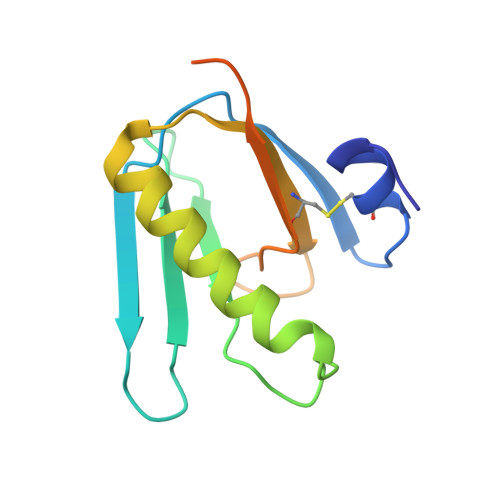
    The self-assembly of proteins into higher order structures is ubiquitous in living systems. It is also an essential process for the bottom-up creation of novel molecular architectures and devices for synthetic biology. However, the complexity of protein-protein interaction surfaces makes it challenging to mimic natural assembly processes in artificial systems. Indeed, many successful computationally designed protein assemblies are prescreened for "designability", limiting the choice of components. Here, we report a simple and pragmatic strategy to assemble chosen multisubunit proteins into more complex structures. A coiled-coil domain appended to one face of the pentameric cholera toxin B-subunit (CTB) enabled the ordered assembly of tubular supra-molecular complexes. Analysis of a tubular structure determined by X-ray crystallography has revealed a hierarchical assembly process that displays features reminiscent of the polymorphic assembly of polyomavirus proteins. The approach provides a simple and straightforward method to direct the assembly of protein building blocks which present either termini on a single face of an oligomer. This scaffolding approach can be used to generate bespoke supramolecular assemblies of functional proteins. Additionally, structural resolution of the scaffolded assemblies highlight "native-state" forced protein-protein interfaces, which may prove useful as starting conformations for future computational design.


  • Organizational Affiliation
    • Astbury Centre for Structural Molecular Biology , University of Leeds , Leeds LS2 9JT , United Kingdom.

Macromolecule Content 

  • Total Structure Weight: 281.39 kDa 
  • Atom Count: 18,049 
  • Modeled Residue Count: 2,237 
  • Deposited Residue Count: 2,480 
  • Unique protein chains: 1

Macromolecules

Find similar proteins by:|  3D Structure
Entity ID: 1
MoleculeChains  Sequence LengthOrganismDetailsImage
Toxin B subunit
A, B, C, D, E
A, B, C, D, E, F, G, H, I, J, K, L, M, N, O, P, Q, R, S, T
124Vibrio choleraeMutation(s): 0 
Gene Names: CTB
UniProt
Find proteins for P01556 (Vibrio cholerae serotype O1 (strain ATCC 39315 / El Tor Inaba N16961))
Explore P01556 
Go to UniProtKB:  P01556
Entity Groups
Sequence Clusters30% Identity50% Identity70% Identity90% Identity95% Identity100% Identity
UniProt GroupP01556
Sequence Annotations
Expand
Reference Sequence

Small Molecules

Ligands 3 Unique
IDChains Name / Formula / InChI Key2D Diagram3D Interactions
MPD

Query on MPD


Download:Ideal Coordinates CCD File
AA [auth D]
AB [auth T]
BA [auth D]
DA [auth G]
EA [auth G]
AA [auth D],
AB [auth T],
BA [auth D],
DA [auth G],
EA [auth G],
FA [auth H],
GA [auth H],
HA [auth H],
JA [auth J],
KA [auth J],
LA [auth L],
MA [auth M],
NA [auth N],
OA [auth O],
PA [auth O],
RA [auth P],
SA [auth P],
UA [auth Q],
V [auth A],
VA [auth R],
W [auth A],
WA [auth R],
X [auth B],
XA [auth R],
Y [auth B],
YA [auth S],
ZA [auth S]
(4S)-2-METHYL-2,4-PENTANEDIOL
C6 H14 O2
SVTBMSDMJJWYQN-YFKPBYRVSA-N
PO4

Query on PO4


Download:Ideal Coordinates CCD File
CA [auth F]
IA [auth I]
QA [auth P]
TA [auth Q]
U [auth A]
CA [auth F],
IA [auth I],
QA [auth P],
TA [auth Q],
U [auth A],
Z [auth D]
PHOSPHATE ION
O4 P
NBIIXXVUZAFLBC-UHFFFAOYSA-K
CL

Query on CL


Download:Ideal Coordinates CCD File
BB [auth T]CHLORIDE ION
Cl
VEXZGXHMUGYJMC-UHFFFAOYSA-M

Experimental Data & Validation

Experimental Data

  • Method: X-RAY DIFFRACTION
  • Resolution: 2.45 Å
  • R-Value Free:  0.214 (Depositor), 0.213 (DCC) 
  • R-Value Work:  0.195 (Depositor), 0.197 (DCC) 
  • R-Value Observed: 0.196 (Depositor) 
Space Group: P 63
Unit Cell:
Length ( Å )Angle ( ˚ )
a = 179.16α = 90
b = 179.16β = 90
c = 192.02γ = 120
Software Package:
Software NamePurpose
REFMACrefinement
PDB_EXTRACTdata extraction
xia2data reduction
Aimlessdata scaling
MOLREPphasing

Structure Validation

View Full Validation Report


Entry History 

& Funding Information

Deposition Data


Funding OrganizationLocationGrant Number
Wellcome TrustUnited Kingdom089308/Z/09/Z
Wellcome TrustUnited Kingdom102572/B/13/Z
Wellcome TrustUnited Kingdom090932/Z/09/Z
Wellcome TrustUnited Kingdom094232/Z/10/Z
Wellcome TrustUnited Kingdom108466/Z/15/Z
Biotechnology and Biological Sciences Research CouncilUnited KingdomBB/M005666/1
German Research FoundationGermanyEXC1074

Revision History  (Full details and data files)

  • Version 1.0: 2019-03-20
    Type: Initial release
  • Version 1.1: 2019-04-10
    Changes: Data collection, Database references
  • Version 1.2: 2019-05-08
    Changes: Advisory, Data collection, Derived calculations
  • Version 1.3: 2019-07-10
    Changes: Data collection
  • Version 1.4: 2024-01-24
    Changes: Data collection, Database references, Refinement description
  • Version 1.5: 2024-11-13
    Changes: Structure summary