6HPS

Near-infrared dual bioluminescence imaging in vivo using infra-luciferin


Experimental Data Snapshot

  • Method: X-RAY DIFFRACTION
  • Resolution: 3.10 Å
  • R-Value Free: 0.333 
  • R-Value Work: 0.277 
  • R-Value Observed: 0.280 

wwPDB Validation   3D Report Full Report


Ligand Structure Quality Assessment 


This is version 1.2 of the entry. See complete history


Literature

Near-infrared dual bioluminescence imaging in mouse models of cancer using infraluciferin.

Stowe, C.L.Burley, T.A.Allan, H.Vinci, M.Kramer-Marek, G.Ciobota, D.M.Parkinson, G.N.Southworth, T.L.Agliardi, G.Hotblack, A.Lythgoe, M.F.Branchini, B.R.Kalber, T.L.Anderson, J.C.Pule, M.A.

(2019) Elife 8

  • DOI: https://doi.org/10.7554/eLife.45801
  • Primary Citation of Related Structures:  
    6HPS

  • PubMed Abstract: 

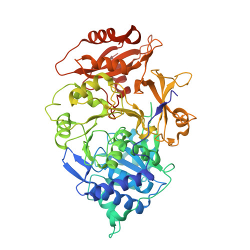
    Bioluminescence imaging (BLI) is ubiquitous in scientific research for the sensitive tracking of biological processes in small animal models. However, due to the attenuation of visible light by tissue, and the limited set of near-infrared bioluminescent enzymes, BLI is largely restricted to monitoring single processes in vivo. Here we show, that by combining stabilised colour mutants of firefly luciferase (FLuc) with the luciferin (LH 2 ) analogue infraluciferin (iLH 2 ), near-infrared dual BLI can be achieved in vivo. The X-ray crystal structure of FLuc with a high-energy intermediate analogue, 5'-O-[N-(dehydroinfraluciferyl)sulfamoyl] adenosine (iDLSA) provides insight into the FLuc-iLH 2 reaction leading to near-infrared light emission. The spectral characterisation and unmixing validation studies reported here established that iLH 2 is superior to LH 2 for the spectral unmixing of bioluminescent signals in vivo; which led to this novel near-infrared dual BLI system being applied to monitor both tumour burden and CAR T cell therapy within a systemically induced mouse tumour model.


  • Organizational Affiliation

    Cancer Institute, University College London, London, United Kingdom.


Macromolecules
Find similar proteins by:  (by identity cutoff)  |  3D Structure
Entity ID: 1
MoleculeChains Sequence LengthOrganismDetailsImage
Luciferin 4-monooxygenase
A, B
543Photinus pyralisMutation(s): 0 
EC: 1.13.12.7
UniProt
Find proteins for P08659 (Photinus pyralis)
Explore P08659 
Go to UniProtKB:  P08659
Entity Groups  
Sequence Clusters30% Identity50% Identity70% Identity90% Identity95% Identity100% Identity
UniProt GroupP08659
Sequence Annotations
Expand
  • Reference Sequence
Small Molecules
Ligands 1 Unique
IDChains Name / Formula / InChI Key2D Diagram3D Interactions
GKH (Subject of Investigation/LOI)
Query on GKH

Download Ideal Coordinates CCD File 
C [auth A],
D [auth B]
[(2~{R},3~{S},4~{R},5~{R})-5-(6-aminopurin-9-yl)-3,4-bis(oxidanyl)oxolan-2-yl]methyl ~{N}-[[2-[(~{E})-2-(6-oxidanyl-1,3-benzothiazol-2-yl)ethenyl]-1,3-thiazol-4-yl]carbonyl]sulfamate
C23 H20 N8 O8 S3
JXXQGJFRVFVJAK-AWGXURSZSA-N
Experimental Data & Validation

Experimental Data

  • Method: X-RAY DIFFRACTION
  • Resolution: 3.10 Å
  • R-Value Free: 0.333 
  • R-Value Work: 0.277 
  • R-Value Observed: 0.280 
  • Space Group: P 1 21 1
Unit Cell:
Length ( Å )Angle ( ˚ )
a = 47.509α = 90
b = 114.016β = 98.18
c = 101.557γ = 90
Software Package:
Software NamePurpose
REFMACrefinement
Aimlessdata scaling
PDB_EXTRACTdata extraction
CrysalisProdata reduction
PHASERphasing

Structure Validation

View Full Validation Report


Ligand Structure Quality Assessment 


Entry History 

Deposition Data

Revision History  (Full details and data files)

  • Version 1.0: 2019-10-23
    Type: Initial release
  • Version 1.1: 2019-10-30
    Changes: Data collection, Database references
  • Version 1.2: 2024-01-24
    Changes: Data collection, Database references, Refinement description