6HOY | pdb_00006hoy

Human Sirt6 in complex with ADP-ribose and the inhibitor trichostatin A


Experimental Data Snapshot

  • Method: X-RAY DIFFRACTION
  • Resolution: 1.70 Å
  • R-Value Free: 
    0.185 (Depositor), 0.185 (DCC) 
  • R-Value Work: 
    0.154 (Depositor), 0.159 (DCC) 
  • R-Value Observed: 
    0.155 (Depositor) 

Starting Model: experimental
View more details

wwPDB Validation 3D Report Full Report

Validation slider image for 6HOY

Ligand Structure Quality Assessment 


This is version 1.2 of the entry. See complete history

Literature

Structural Basis of Sirtuin 6 Inhibition by the Hydroxamate Trichostatin A: Implications for Protein Deacylase Drug Development.

You, W.Steegborn, C.

(2018) J Med Chem 61: 10922-10928

  • DOI: https://doi.org/10.1021/acs.jmedchem.8b01455
  • Primary Citation Related Structures: 
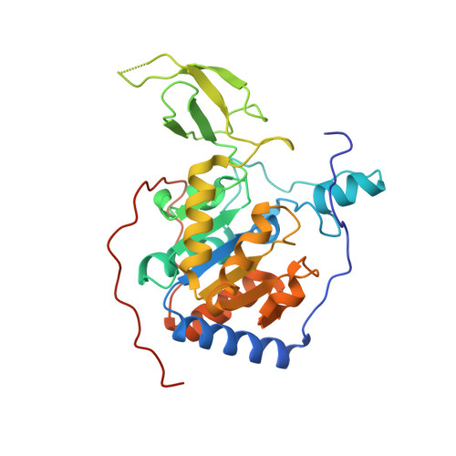
    6HOY

  • PubMed Abstract: 

    Protein lysine deacylases comprise three zinc-dependent families and the NAD + -dependent sirtuins Sirt1-7, which contribute to aging-related diseases. Few Sirt6-specific inhibitors are available. Trichostatin A, which belongs to the potent, zinc-chelating hydroxamate inhibitors of zinc-dependent deacylases, was recently found to potently and isoform-specifically inhibit Sirt6. We solved a crystal structure of a Sirt6/ADP-ribose/trichostatin A complex, which reveals nicotinamide pocket and acyl channel as binding site and provides interaction details supporting the development of improved deacylase inhibitors.


  • Organizational Affiliation
    • Department of Biochemistry , University of Bayreuth , Universitätsstraße 30 , 95445 Bayreuth , Germany.

Macromolecule Content 

  • Total Structure Weight: 70.36 kDa 
  • Atom Count: 4,902 
  • Modeled Residue Count: 557 
  • Deposited Residue Count: 604 
  • Unique protein chains: 1

Macromolecules

Find similar proteins by:|  3D Structure
Entity ID: 1
MoleculeChains  Sequence LengthOrganismDetailsImage
NAD-dependent protein deacetylase sirtuin-6
A, B
302Homo sapiensMutation(s): 0 
Gene Names: SIRT6SIR2L6
EC: 3.5.1 (PDB Primary Data), 2.4.2 (UniProt), 2.3.1 (UniProt), 2.3.1.286 (UniProt)
UniProt & NIH Common Fund Data Resources
Find proteins for Q8N6T7 (Homo sapiens)
Explore Q8N6T7 
Go to UniProtKB:  Q8N6T7
PHAROS:  Q8N6T7
GTEx:  ENSG00000077463 
Entity Groups
Sequence Clusters30% Identity50% Identity70% Identity90% Identity95% Identity100% Identity
UniProt GroupQ8N6T7
Sequence Annotations
Expand
Reference Sequence

Small Molecules

Ligands 6 Unique
IDChains Name / Formula / InChI Key2D Diagram3D Interactions
AR6

Query on AR6


Download:Ideal Coordinates CCD File
C [auth A],
M [auth B]
[(2R,3S,4R,5R)-5-(6-AMINOPURIN-9-YL)-3,4-DIHYDROXY-OXOLAN-2-YL]METHYL[HYDROXY-[[(2R,3S,4R,5S)-3,4,5-TRIHYDROXYOXOLAN-2-YL]METHOXY]PHOSPHORYL] HYDROGEN PHOSPHATE
C15 H23 N5 O14 P2
SRNWOUGRCWSEMX-ZQSHOCFMSA-N
TSN

Query on TSN


Download:Ideal Coordinates CCD File
K [auth A],
T [auth B]
TRICHOSTATIN A
C17 H22 N2 O3
RTKIYFITIVXBLE-QEQCGCAPSA-N
PG4

Query on PG4


Download:Ideal Coordinates CCD File
E [auth A],
O [auth B]
TETRAETHYLENE GLYCOL
C8 H18 O5
UWHCKJMYHZGTIT-UHFFFAOYSA-N
SO4

Query on SO4


Download:Ideal Coordinates CCD File
G [auth A]
H [auth A]
I [auth A]
J [auth A]
Q [auth B]
G [auth A],
H [auth A],
I [auth A],
J [auth A],
Q [auth B],
R [auth B],
S [auth B]
SULFATE ION
O4 S
QAOWNCQODCNURD-UHFFFAOYSA-L
ZN

Query on ZN


Download:Ideal Coordinates CCD File
D [auth A],
N [auth B]
ZINC ION
Zn
PTFCDOFLOPIGGS-UHFFFAOYSA-N
EDO

Query on EDO


Download:Ideal Coordinates CCD File
F [auth A],
L [auth B],
P [auth B]
1,2-ETHANEDIOL
C2 H6 O2
LYCAIKOWRPUZTN-UHFFFAOYSA-N

Experimental Data & Validation

Experimental Data

  • Method: X-RAY DIFFRACTION
  • Resolution: 1.70 Å
  • R-Value Free:  0.185 (Depositor), 0.185 (DCC) 
  • R-Value Work:  0.154 (Depositor), 0.159 (DCC) 
  • R-Value Observed: 0.155 (Depositor) 
Space Group: P 63
Unit Cell:
Length ( Å )Angle ( ˚ )
a = 91.291α = 90
b = 91.291β = 90
c = 143.681γ = 120
Software Package:
Software NamePurpose
REFMACrefinement
XDSdata reduction
XDSdata scaling
PHASERphasing

Structure Validation

View Full Validation Report


Ligand Structure Quality Assessment 


Entry History 

Deposition Data

Revision History  (Full details and data files)

  • Version 1.0: 2018-11-14
    Type: Initial release
  • Version 1.1: 2019-04-24
    Changes: Data collection, Database references
  • Version 1.2: 2024-01-24
    Changes: Data collection, Database references, Refinement description, Structure summary