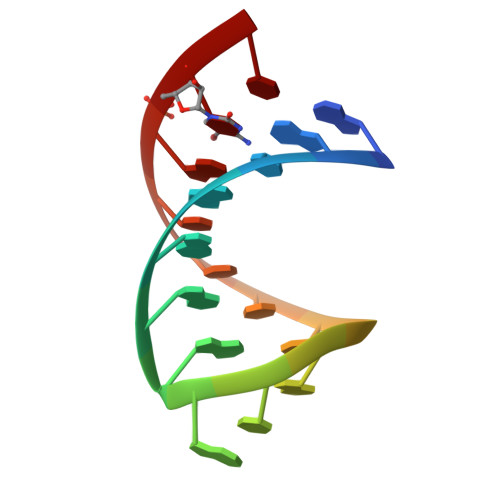
6HC5

The structure of the G. violaceus guanidine II riboswitch P2 stem-loop with audouine


Experimental Data Snapshot

  • Method: X-RAY DIFFRACTION
  • Resolution: 1.41 Å
  • R-Value Free: 0.199 
  • R-Value Work: 0.175 
  • R-Value Observed: 0.176 

wwPDB Validation   3D Report Full Report


This is version 1.2 of the entry. See complete history


Literature

Structure-guided design of a high-affinity ligand for a riboswitch.

Huang, L.Wang, J.Wilson, T.J.Lilley, D.M.J.

(2019) RNA 25: 423-430

  • DOI: https://doi.org/10.1261/rna.069567.118
  • Primary Citation of Related Structures:  
    6HBT, 6HBX, 6HC5

  • PubMed Abstract: 

    We have designed structure-based ligands for the guanidine-II riboswitch that bind with enhanced affinity, exploiting the twin binding sites created by loop-loop interaction. We synthesized diguanidine species, comprising two guanidino groups covalently connected by C n linkers where n = 4 or 5. Calorimetric and fluorescent analysis shows that these ligands bind with a 10-fold higher affinity to the riboswitch compared to guanidine. We determined X-ray crystal structures of the riboswitch bound to the new ligands, showing that the guanidino groups are bound to both nucleobases and backbone within the binding pockets, analogously to guanidine binding. The connecting chain passes through side openings in the binding pocket and traverses the minor groove of the RNA. The combination of the riboswitch loop-loop interaction and our novel ligands has potential applications in chemical biology.


  • Organizational Affiliation

    Cancer Research UK Nucleic Acid Structure Research Group, MSI/WTB Complex, The University of Dundee, Dundee DD1 5EH, United Kingdom.


Macromolecules

Find similar nucleic acids by:  Sequence   |   3D Structure  

Entity ID: 1
MoleculeChains LengthOrganismImage
RNA (5'-R(*GP*GP*UP*GP*GP*GP*GP*AP*CP*GP*AP*CP*CP*CP*CP*AP*(CBV)P*C)-3')18Gloeobacter violaceus
Sequence Annotations
Expand
  • Reference Sequence
Experimental Data & Validation

Experimental Data

  • Method: X-RAY DIFFRACTION
  • Resolution: 1.41 Å
  • R-Value Free: 0.199 
  • R-Value Work: 0.175 
  • R-Value Observed: 0.176 
  • Space Group: H 3 2
Unit Cell:
Length ( Å )Angle ( ˚ )
a = 55.826α = 90
b = 55.826β = 90
c = 133.363γ = 120
Software Package:
Software NamePurpose
PHENIXrefinement
xia2data reduction
xia2data scaling
PHENIXphasing

Structure Validation

View Full Validation Report


Entry History & Funding Information

Deposition Data


Funding OrganizationLocationGrant Number
Cancer Research UKUnited KingdomA18604

Revision History  (Full details and data files)

  • Version 1.0: 2019-02-20
    Type: Initial release
  • Version 1.1: 2019-03-27
    Changes: Data collection, Database references
  • Version 1.2: 2024-05-15
    Changes: Data collection, Database references