Structural and Functional Analyses of the Human PDH Complex Suggest a "Division-of-Labor" Mechanism by Local E1 and E3 Clusters.
Prajapati, S., Haselbach, D., Wittig, S., Patel, M.S., Chari, A., Schmidt, C., Stark, H., Tittmann, K.(2019) Structure 27: 1124-1136.e4
- PubMed: 31130485 Search on PubMed
- DOI: https://doi.org/10.1016/j.str.2019.04.009
- Primary Citation Related Structures:
6H55, 6H60 - PubMed Abstract:
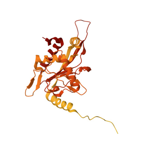
The pseudo-atomic structural model of human pyruvate dehydrogenase complex (PDHc) core composed of full-length E2 and E3BP components, calculated from our cryoelectron microscopy-derived density maps at 6-Å resolution, is similar to those of prokaryotic E2 structures. The spatial organization of human PDHc components as evidenced by negative-staining electron microscopy and native mass spectrometry is not homogeneous, and entails the unanticipated formation of local clusters of E1:E2 and E3BP:E3 complexes. Such uneven, clustered organization translates into specific duties for E1-E2 clusters (oxidative decarboxylation and acetyl transfer) and E3BP-E3 clusters (regeneration of reduced lipoamide) corresponding to half-reactions of the PDHc catalytic cycle. The addition of substrate coenzyme A modulates the conformational landscape of PDHc, in particular of the lipoyl domains, extending the postulated multiple random coupling mechanism. The conformational and associated chemical landscapes of PDHc are thus not determined entirely stochastically, but are restrained and channeled through an asymmetric architecture and further modulated by substrate binding.
- Department of Molecular Enzymology, Göttingen Centre for Molecular Biosciences and Albrecht-von-Haller Institute, Georg-August University Göttingen, Julia-Lermontowa-Weg 3, 37077 Göttingen, Germany; Department of Structural Dynamics, Max-Planck-Institute for Biophysical Chemistry Göttingen, Am Fassberg 11, 37077 Göttingen, Germany.
Organizational Affiliation: