Crystal structure of carbonic anhydrase CaNce103p from the pathogenic yeast Candida albicans.
Dostal, J., Brynda, J., Blaha, J., Machacek, S., Heidingsfeld, O., Pichova, I.(2018) BMC Struct Biol 18: 14-14
- PubMed: 30367660 Search on PubMedSearch on PubMed Central
- DOI: https://doi.org/10.1186/s12900-018-0093-4
- Primary Citation Related Structures:
6GWU - PubMed Abstract:
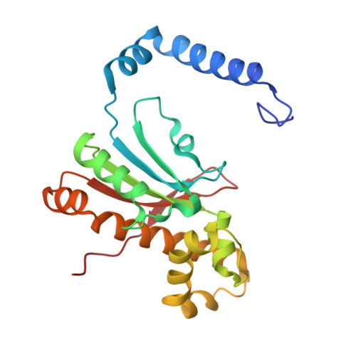
The pathogenic yeast Candida albicans can proliferate in environments with different carbon dioxide concentrations thanks to the carbonic anhydrase CaNce103p, which accelerates spontaneous conversion of carbon dioxide to bicarbonate and vice versa. Without functional CaNce103p, C. albicans cannot survive in atmospheric air. CaNce103p falls into the β-carbonic anhydrase class, along with its ortholog ScNce103p from Saccharomyces cerevisiae. The crystal structure of CaNce103p is of interest because this enzyme is a potential target for surface disinfectants. Recombinant CaNce103p was prepared in E. coli, and its crystal structure was determined at 2.2 Å resolution. CaNce103p forms a homotetramer organized as a dimer of dimers, in which the dimerization and tetramerization surfaces are perpendicular. Although the physiological role of CaNce103p is similar to that of ScNce103p from baker's yeast, on the structural level it more closely resembles carbonic anhydrase from the saprophytic fungus Sordaria macrospora, which is also tetrameric. Dimerization is mediated by two helices in the N-terminal domain of the subunits. The N-terminus of CaNce103p is flexible, and crystals were obtained only upon truncation of the first 29 amino acids. Analysis of CaNce103p variants truncated by 29, 48 and 61 amino acids showed that residues 30-48 are essential for dimerization. Each subunit contains a zinc atom in the active site and displays features characteristic of type I β-carbonic anhydrases. Zinc is tetrahedrally coordinated by one histidine residue, two cysteine residues and a molecule of β-mercaptoethanol originating from the crystallization buffer. The active sites are accessible via substrate tunnels, which are slightly longer and narrower than those observed in other fungal carbonic anhydrases. CaNce103p is a β-class homotetrameric metalloenzyme composed of two homodimers. Its structure closely resembles those of other β-type carbonic anhydrases, in particular CAS1 from Sordaria macrospora. The main differences occur in the N-terminal part and the substrate tunnel. Detailed knowledge of the CaNce103p structure and the properties of the substrate tunnel in particular will facilitate design of selective inhibitors of this enzyme.
- Institute of Organic Chemistry and Biochemistry of the Czech Academy of Sciences, Flemingovo náměstí 2, 166 10, Prague, Czech Republic.
Organizational Affiliation: