Solvent Networks Tune Thermodynamics of Oligosaccharide Complex Formation in an Extended Protein Binding Site.
Kunstmann, S., Gohlke, U., Broeker, N.K., Roske, Y., Heinemann, U., Santer, M., Barbirz, S.(2018) J Am Chem Soc 140: 10447-10455
- PubMed: 30044908 Search on PubMed
- DOI: https://doi.org/10.1021/jacs.8b03719
- Primary Citation Related Structures:
6G0X, 6GVP, 6GVR - PubMed Abstract:
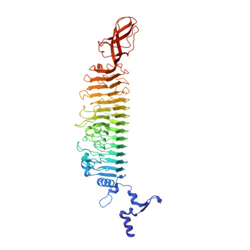
The principles of protein-glycan binding are still not well understood on a molecular level. Attempts to link affinity and specificity of glycan recognition to structure suffer from the general lack of model systems for experimental studies and the difficulty to describe the influence of solvent. We have experimentally and computationally addressed energetic contributions of solvent in protein-glycan complex formation in the tailspike protein (TSP) of E. coli bacteriophage HK620. HK620TSP is a 230 kDa native trimer of right-handed, parallel beta-helices that provide extended, rigid binding sites for bacterial cell surface O-antigen polysaccharides. A set of high-affinity mutants bound hexa- or pentasaccharide O-antigen fragments with very similar affinities even though hexasaccharides introduce an additional glucose branch into an occluded protein surface cavity. Remarkably different thermodynamic binding signatures were found for different mutants; however, crystal structure analyses indicated that no major oligosaccharide or protein topology changes had occurred upon complex formation. This pointed to a solvent effect. Molecular dynamics simulations using a mobility-based approach revealed an extended network of solvent positions distributed over the entire oligosaccharide binding site. However, free energy calculations showed that a small water network inside the glucose-binding cavity had the most notable influence on the thermodynamic signature. The energy needed to displace water from the glucose binding pocket depended on the amino acid at the entrance, in agreement with the different amounts of enthalpy-entropy compensation found for introducing glucose into the pocket in the different mutants. Studies with small molecule drugs have shown before that a few active water molecules can control protein complex formation. HK620TSP oligosaccharide binding shows that similar fundamental principles also apply for glycans, where a small number of water molecules can dominate the thermodynamic signature in an extended binding site.
- Physikalische Biochemie , Universität Potsdam , Karl-Liebknecht-Str. 24-25 , 14476 Potsdam , Germany.
Organizational Affiliation: