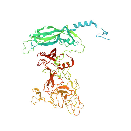
6GSH

Feline Calicivirus Strain F9

  • Classification: VIRUS
  • Organism(s): Feline calicivirus
  • Expression System: Felis catus
  • Mutation(s): No 

  • Deposited: 2018-06-14 Released: 2019-01-16 
  • Deposition Author(s): Conley, M.J., Bhella, D.
  • Funding Organization(s): Medical Research Council (United Kingdom), Biotechnology and Biological Sciences Research Council

Experimental Data Snapshot

  • Method: ELECTRON MICROSCOPY
  • Resolution: 3.00 Å
  • Aggregation State: PARTICLE 
  • Reconstruction Method: SINGLE PARTICLE 

wwPDB Validation   3D Report Full Report


This is version 1.4 of the entry. See complete history


Literature

Calicivirus VP2 forms a portal-like assembly following receptor engagement.

Conley, M.J.McElwee, M.Azmi, L.Gabrielsen, M.Byron, O.Goodfellow, I.G.Bhella, D.

(2019) Nature 565: 377-381

  • DOI: https://doi.org/10.1038/s41586-018-0852-1
  • Primary Citation of Related Structures:  
    6GSH, 6GSI

  • PubMed Abstract: 
  • To initiate infection, many viruses enter their host cells by triggering endocytosis following receptor engagement. However, the mechanisms by which non-enveloped viruses escape the endosome are poorly understood. Here we present near-atomic-resolution cryo-electron microscopy structures for feline calicivirus both undecorated and labelled with a soluble fragment of its cellular receptor, feline junctional adhesion molecule A ...

    To initiate infection, many viruses enter their host cells by triggering endocytosis following receptor engagement. However, the mechanisms by which non-enveloped viruses escape the endosome are poorly understood. Here we present near-atomic-resolution cryo-electron microscopy structures for feline calicivirus both undecorated and labelled with a soluble fragment of its cellular receptor, feline junctional adhesion molecule A. We show that VP2, a minor capsid protein encoded by all caliciviruses 1,2 , forms a large portal-like assembly at a unique three-fold axis of symmetry, following receptor engagement. This assembly-which was not detected in undecorated virions-is formed of twelve copies of VP2, arranged with their hydrophobic N termini pointing away from the virion surface. Local rearrangement at the portal site leads to the opening of a pore in the capsid shell. We hypothesize that the portal-like assembly functions as a channel for the delivery of the calicivirus genome, through the endosomal membrane, into the cytoplasm of a host cell, thereby initiating infection. VP2 was previously known to be critical for the production of infectious virus 3 ; our findings provide insights into its structure and function that advance our understanding of the Caliciviridae.


    Organizational Affiliation

    Medical Research Council University of Glasgow Centre for Virus Research, Glasgow, UK. david.bhella@glasgow.ac.uk.


Macromolecules
Find similar proteins by:  (by identity cutoff)  |  3D Structure
Entity ID: 1
MoleculeChainsSequence LengthOrganismDetailsImage
VP1
A, B, C
669Feline calicivirusMutation(s): 0 
UniProt
Find proteins for A2T4P8 (Feline calicivirus)
Explore A2T4P8 
Go to UniProtKB:  A2T4P8
Entity Groups  
Sequence Clusters30% Identity50% Identity70% Identity90% Identity95% Identity100% Identity
UniProt GroupA2T4P8
Protein Feature View
Expand
  • Reference Sequence
Small Molecules
Ligands 1 Unique
IDChainsName / Formula / InChI Key2D Diagram3D Interactions
K
Query on K

Download Ideal Coordinates CCD File 
D [auth A],
E [auth B],
F [auth C]
POTASSIUM ION
K
NPYPAHLBTDXSSS-UHFFFAOYSA-N
 Ligand Interaction
Experimental Data & Validation

Experimental Data

  • Method: ELECTRON MICROSCOPY
  • Resolution: 3.00 Å
  • Aggregation State: PARTICLE 
  • Reconstruction Method: SINGLE PARTICLE 
EM Software:
TaskSoftware PackageVersion
RECONSTRUCTIONRELION2.1

Structure Validation

View Full Validation Report


Entry History & Funding Information

Deposition Data


Funding OrganizationLocationGrant Number
Medical Research Council (United Kingdom)United KingdomMC_UU_12014/7
Biotechnology and Biological Sciences Research CouncilUnited KingdomBB/J013854/1

Revision History  (Full details and data files)

  • Version 1.0: 2019-01-16
    Type: Initial release
  • Version 1.1: 2019-01-23
    Changes: Data collection, Database references
  • Version 1.2: 2019-01-30
    Changes: Data collection, Database references
  • Version 1.3: 2019-02-06
    Changes: Data collection, Derived calculations
  • Version 1.4: 2019-12-11
    Changes: Other