6GRJ

Structure of the AhlB pore of the tripartite alpha-pore forming toxin, AHL, from Aeromonas hydrophila.


Experimental Data Snapshot

  • Method: X-RAY DIFFRACTION
  • Resolution: 2.94 Å
  • R-Value Free: 0.239 
  • R-Value Work: 0.222 
  • R-Value Observed: 0.223 

wwPDB Validation   3D Report Full Report


This is version 1.2 of the entry. See complete history


Literature

Identification and structural analysis of the tripartite alpha-pore forming toxin of Aeromonas hydrophila.

Wilson, J.S.Churchill-Angus, A.M.Davies, S.P.Sedelnikova, S.E.Tzokov, S.B.Rafferty, J.B.Bullough, P.A.Bisson, C.Baker, P.J.

(2019) Nat Commun 10: 2900-2900

  • DOI: https://doi.org/10.1038/s41467-019-10777-x
  • Primary Citation of Related Structures:  
    6GRJ, 6GRK, 6H2D, 6H2E, 6H2F, 6R1J

  • PubMed Abstract: 

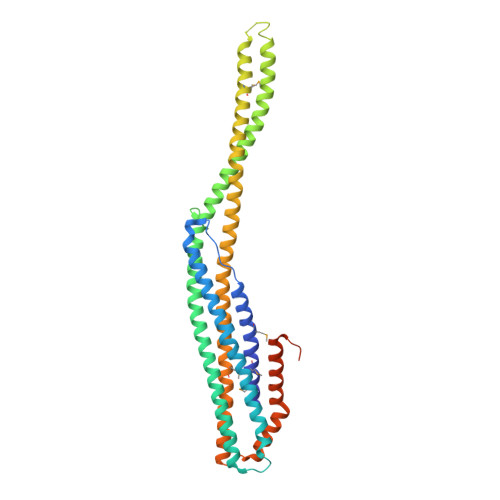
    The alpha helical CytolysinA family of pore forming toxins (α-PFT) contains single, two, and three component members. Structures of the single component Eschericia coli ClyA and the two component Yersinia enterolytica YaxAB show both undergo conformational changes from soluble to pore forms, and oligomerization to produce the active pore. Here we identify tripartite α-PFTs in pathogenic Gram negative bacteria, including Aeromonas hydrophila (AhlABC). We show that the AhlABC toxin requires all three components for maximal cell lysis. We present structures of pore components which describe a bi-fold hinge mechanism for soluble to pore transition in AhlB and a contrasting tetrameric assembly employed by soluble AhlC to hide their hydrophobic membrane associated residues. We propose a model of pore assembly where the AhlC tetramer dissociates, binds a single membrane leaflet, recruits AhlB promoting soluble to pore transition, prior to AhlA binding to form the active hydrophilic lined pore.


  • Organizational Affiliation

    Department of Molecular Biology and Biotechnology, University of Sheffield, Firth Court, Western Bank, Sheffield, South Yorkshire, S10 2TN, UK.


Macromolecules
Find similar proteins by:  (by identity cutoff)  |  3D Structure
Entity ID: 1
MoleculeChains Sequence LengthOrganismDetailsImage
AhlB367Aeromonas hydrophilaMutation(s): 1 
Gene Names: A9R12_16795
Membrane Entity: Yes 
UniProt
Find proteins for A0A454GEH4 (Aeromonas hydrophila)
Explore A0A454GEH4 
Go to UniProtKB:  A0A454GEH4
Entity Groups  
Sequence Clusters30% Identity50% Identity70% Identity90% Identity95% Identity100% Identity
UniProt GroupA0A454GEH4
Sequence Annotations
Expand
  • Reference Sequence
Small Molecules
Ligands 4 Unique
IDChains Name / Formula / InChI Key2D Diagram3D Interactions
MPD
Query on MPD

Download Ideal Coordinates CCD File 
AA [auth C]
AB [auth H]
BA [auth C]
BB [auth H]
GB [auth I]
AA [auth C],
AB [auth H],
BA [auth C],
BB [auth H],
GB [auth I],
K [auth G],
L [auth G],
M [auth G],
NA [auth E],
OA [auth E],
R [auth J],
S [auth J],
UA [auth F],
V [auth A]
(4S)-2-METHYL-2,4-PENTANEDIOL
C6 H14 O2
SVTBMSDMJJWYQN-YFKPBYRVSA-N
PO4
Query on PO4

Download Ideal Coordinates CCD File 
N [auth G],
PA [auth E]
PHOSPHATE ION
O4 P
NBIIXXVUZAFLBC-UHFFFAOYSA-K
CL
Query on CL

Download Ideal Coordinates CCD File 
CA [auth C]
CB [auth H]
DA [auth C]
DB [auth H]
EA [auth C]
CA [auth C],
CB [auth H],
DA [auth C],
DB [auth H],
EA [auth C],
EB [auth H],
FA [auth C],
GA [auth C],
HB [auth I],
IB [auth I],
JB [auth I],
KA [auth D],
KB [auth I],
LA [auth D],
O [auth G],
P [auth G],
Q [auth G],
QA [auth E],
RA [auth E],
T [auth J],
VA [auth F],
W [auth A],
WA [auth F],
X [auth B],
XA [auth F],
Y [auth B],
YA [auth F]
CHLORIDE ION
Cl
VEXZGXHMUGYJMC-UHFFFAOYSA-M
NA
Query on NA

Download Ideal Coordinates CCD File 
FB [auth H]
HA [auth C]
IA [auth C]
JA [auth C]
MA [auth D]
FB [auth H],
HA [auth C],
IA [auth C],
JA [auth C],
MA [auth D],
SA [auth E],
TA [auth E],
U [auth J],
Z [auth B],
ZA [auth F]
SODIUM ION
Na
FKNQFGJONOIPTF-UHFFFAOYSA-N
Modified Residues  1 Unique
IDChains TypeFormula2D DiagramParent
MSE
Query on MSE
A [auth G]
B [auth J]
C [auth A]
D [auth B]
E [auth C]
A [auth G],
B [auth J],
C [auth A],
D [auth B],
E [auth C],
F [auth D],
G [auth E],
H [auth F],
I [auth H],
J [auth I]
L-PEPTIDE LINKINGC5 H11 N O2 SeMET
Experimental Data & Validation

Experimental Data

  • Method: X-RAY DIFFRACTION
  • Resolution: 2.94 Å
  • R-Value Free: 0.239 
  • R-Value Work: 0.222 
  • R-Value Observed: 0.223 
  • Space Group: C 1 2 1
Unit Cell:
Length ( Å )Angle ( ˚ )
a = 363.644α = 90
b = 116.526β = 118.01
c = 217.409γ = 90
Software Package:
Software NamePurpose
REFMACrefinement
DIALSdata reduction
DIALSdata scaling
SHELXDEphasing

Structure Validation

View Full Validation Report


Entry History 

Deposition Data

Revision History  (Full details and data files)

  • Version 1.0: 2019-07-03
    Type: Initial release
  • Version 1.1: 2019-07-10
    Changes: Data collection, Database references
  • Version 1.2: 2019-07-17
    Changes: Data collection, Database references