6GRE

Crystal structure of the tandem DUF26 ectodomain from the Arabidopsis thaliana cysteine-rich receptor-like protein PDLP5.


Experimental Data Snapshot

  • Method: X-RAY DIFFRACTION
  • Resolution: 1.29 Å
  • R-Value Free: 0.210 
  • R-Value Work: 0.186 
  • R-Value Observed: 0.188 

wwPDB Validation   3D Report Full Report


Ligand Structure Quality Assessment 


This is version 2.0 of the entry. See complete history


Literature

Mechanistic insights into the evolution of DUF26-containing proteins in land plants.

Vaattovaara, A.Brandt, B.Rajaraman, S.Safronov, O.Veidenberg, A.Luklova, M.Kangasjarvi, J.Loytynoja, A.Hothorn, M.Salojarvi, J.Wrzaczek, M.

(2019) Commun Biol 2: 56-56

  • DOI: https://doi.org/10.1038/s42003-019-0306-9
  • Primary Citation of Related Structures:  
    6GRE, 6GRF

  • PubMed Abstract: 

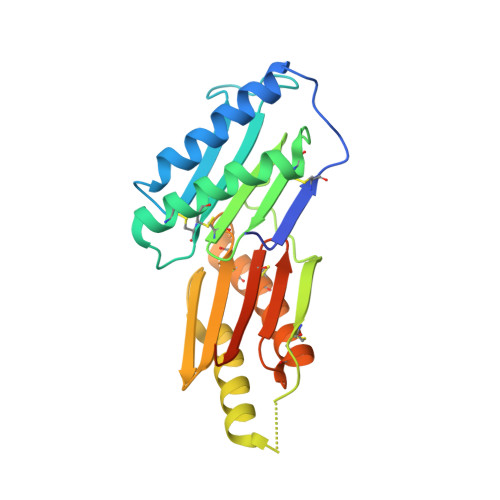
    Large protein families are a prominent feature of plant genomes and their size variation is a key element for adaptation. However, gene and genome duplications pose difficulties for functional characterization and translational research. Here we infer the evolutionary history of the DOMAIN OF UNKNOWN FUNCTION (DUF) 26-containing proteins. The DUF26 emerged in secreted proteins. Domain duplications and rearrangements led to the appearance of CYSTEINE-RICH RECEPTOR-LIKE PROTEIN KINASES (CRKs) and PLASMODESMATA-LOCALIZED PROTEINS (PDLPs). The DUF26 is land plant-specific but structural analyses of PDLP ectodomains revealed strong similarity to fungal lectins and thus may constitute a group of plant carbohydrate-binding proteins. CRKs expanded through tandem duplications and preferential retention of duplicates following whole genome duplications, whereas PDLPs evolved according to the dosage balance hypothesis. We propose that new gene families mainly expand through small-scale duplications, while fractionation and genetic drift after whole genome multiplications drive families towards dosage balance.


  • Organizational Affiliation

    1Organismal and Evolutionary Biology Research Programme, Viikki Plant Science Centre, VIPS, Faculty of Biological and Environmental Sciences, University of Helsinki, Viikinkaari 1 (POB65), FI-00014 Helsinki, Finland.


Macromolecules
Find similar proteins by:  (by identity cutoff)  |  3D Structure
Entity ID: 1
MoleculeChains Sequence LengthOrganismDetailsImage
Cysteine-rich repeat secretory protein 2
A, B
228Arabidopsis thalianaMutation(s): 0 
Gene Names: CRRSP2HWI1At1g70690F5A18.13
UniProt
Find proteins for Q8GUJ2 (Arabidopsis thaliana)
Explore Q8GUJ2 
Go to UniProtKB:  Q8GUJ2
Entity Groups  
Sequence Clusters30% Identity50% Identity70% Identity90% Identity95% Identity100% Identity
UniProt GroupQ8GUJ2
Sequence Annotations
Expand
  • Reference Sequence
Oligosaccharides

Help

Entity ID: 2
MoleculeChains Length2D Diagram Glycosylation3D Interactions
2-acetamido-2-deoxy-beta-D-glucopyranose-(1-4)-2-acetamido-2-deoxy-beta-D-glucopyranose
C, D, E
2N-Glycosylation
Glycosylation Resources
GlyTouCan:  G42666HT
GlyCosmos:  G42666HT
GlyGen:  G42666HT
Small Molecules
Ligands 6 Unique
IDChains Name / Formula / InChI Key2D Diagram3D Interactions
NAG
Query on NAG

Download Ideal Coordinates CCD File 
F [auth A]2-acetamido-2-deoxy-beta-D-glucopyranose
C8 H15 N O6
OVRNDRQMDRJTHS-FMDGEEDCSA-N
AE3
Query on AE3

Download Ideal Coordinates CCD File 
R [auth B]2-(2-ETHOXYETHOXY)ETHANOL
C6 H14 O3
XXJWXESWEXIICW-UHFFFAOYSA-N
SO4
Query on SO4

Download Ideal Coordinates CCD File 
L [auth A],
S [auth B]
SULFATE ION
O4 S
QAOWNCQODCNURD-UHFFFAOYSA-L
GOL
Query on GOL

Download Ideal Coordinates CCD File 
P [auth B]GLYCEROL
C3 H8 O3
PEDCQBHIVMGVHV-UHFFFAOYSA-N
EDO
Query on EDO

Download Ideal Coordinates CCD File 
G [auth A]
H [auth A]
I [auth A]
J [auth A]
M [auth B]
G [auth A],
H [auth A],
I [auth A],
J [auth A],
M [auth B],
N [auth B],
O [auth B]
1,2-ETHANEDIOL
C2 H6 O2
LYCAIKOWRPUZTN-UHFFFAOYSA-N
NA
Query on NA

Download Ideal Coordinates CCD File 
K [auth A],
Q [auth B]
SODIUM ION
Na
FKNQFGJONOIPTF-UHFFFAOYSA-N
Experimental Data & Validation

Experimental Data

  • Method: X-RAY DIFFRACTION
  • Resolution: 1.29 Å
  • R-Value Free: 0.210 
  • R-Value Work: 0.186 
  • R-Value Observed: 0.188 
  • Space Group: P 1
Unit Cell:
Length ( Å )Angle ( ˚ )
a = 41.76α = 97.7
b = 47.97β = 102.72
c = 62.19γ = 99.86
Software Package:
Software NamePurpose
REFMACrefinement
XDSdata reduction
XSCALEdata scaling
SHARPphasing

Structure Validation

View Full Validation Report


Ligand Structure Quality Assessment 


Entry History & Funding Information

Deposition Data


Funding OrganizationLocationGrant Number
Swiss National Science FoundationSwitzerland31003A_176237

Revision History  (Full details and data files)

  • Version 1.0: 2018-12-26
    Type: Initial release
  • Version 1.1: 2019-02-27
    Changes: Data collection, Database references
  • Version 2.0: 2020-07-29
    Type: Remediation
    Reason: Carbohydrate remediation
    Changes: Atomic model, Data collection, Derived calculations, Structure summary