6GQ5

Crystal Structure of the PSMalpha3 Peptide Mutant L15A Forming Cross-Alpha Amyloid-like Fibril


Experimental Data Snapshot

  • Method: X-RAY DIFFRACTION
  • Resolution: 1.50 Å
  • R-Value Free: 0.245 
  • R-Value Work: 0.194 
  • R-Value Observed: 0.200 

wwPDB Validation   3D Report Full Report


This is version 1.3 of the entry. See complete history


Literature

Staphylococcus aureus PSM alpha 3 Cross-alpha Fibril Polymorphism and Determinants of Cytotoxicity.

Tayeb-Fligelman, E.Salinas, N.Tabachnikov, O.Landau, M.

(2020) Structure 28: 301-313.e6

  • DOI: https://doi.org/10.1016/j.str.2019.12.006
  • Primary Citation of Related Structures:  
    6GQ2, 6GQ5, 6GQC

  • PubMed Abstract: 

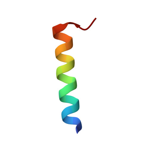
    The phenol-soluble modulin (PSM) peptide family, secreted by Staphylococcus aureus, performs various virulence activities, some mediated by the formation of amyloid fibrils of diverse architectures. Specifically, PSMα1 and PSMα4 structure the S. aureus biofilm by assembling into robust cross-β amyloid fibrils. PSMα3, the most cytotoxic member of the family, assembles into cross-α fibrils in which α helices stack into tightly mated sheets, mimicking the cross-β architecture. Here we demonstrate that massive T cell deformation and death are linked with PSMα3 aggregation and co-localization with cell membranes. Our extensive mutagenesis analyses support the role of positive charges, and especially Lys17, in interactions with the membrane and suggest their regulation by inter- and intra-helical electrostatic interactions within the cross-α fibril. We hypothesize that PSMα3 cytotoxicity is governed by the ability to form cross-α fibrils and involves a dynamic process of co-aggregation with the cell membrane, rupturing it.


  • Organizational Affiliation

    Department of Biology, Technion-Israel Institute of Technology, Haifa 3200003, Israel.


Macromolecules

Find similar proteins by:  Sequence   |   3D Structure  

Entity ID: 1
MoleculeChains Sequence LengthOrganismDetailsImage
Phenol-soluble modulin alpha 3 peptide22Staphylococcus aureus RF122Mutation(s): 1 
UniProt
Find proteins for P0C807 (Staphylococcus aureus (strain bovine RF122 / ET3-1))
Explore P0C807 
Go to UniProtKB:  P0C807
Entity Groups  
Sequence Clusters30% Identity50% Identity70% Identity90% Identity95% Identity100% Identity
UniProt GroupP0C807
Sequence Annotations
Expand
  • Reference Sequence
Small Molecules
Ligands 1 Unique
IDChains Name / Formula / InChI Key2D Diagram3D Interactions
MRD
Query on MRD

Download Ideal Coordinates CCD File 
B [auth A](4R)-2-METHYLPENTANE-2,4-DIOL
C6 H14 O2
SVTBMSDMJJWYQN-RXMQYKEDSA-N
Experimental Data & Validation

Experimental Data

  • Method: X-RAY DIFFRACTION
  • Resolution: 1.50 Å
  • R-Value Free: 0.245 
  • R-Value Work: 0.194 
  • R-Value Observed: 0.200 
  • Space Group: P 1 21 1
Unit Cell:
Length ( Å )Angle ( ˚ )
a = 28.41α = 90
b = 10.67β = 105.87
c = 29.6γ = 90
Software Package:
Software NamePurpose
REFMACrefinement
XDSdata reduction
XSCALEdata scaling
PHASERphasing
PDB_EXTRACTdata extraction

Structure Validation

View Full Validation Report


Entry History 

Deposition Data

Revision History  (Full details and data files)

  • Version 1.0: 2019-06-19
    Type: Initial release
  • Version 1.1: 2019-12-25
    Changes: Data collection
  • Version 1.2: 2020-07-01
    Changes: Database references
  • Version 1.3: 2024-01-17
    Changes: Data collection, Database references, Refinement description