6GIS

Structural basis of human clamp sliding on DNA


Experimental Data Snapshot

  • Method: X-RAY DIFFRACTION
  • Resolution: 2.82 Å
  • R-Value Free: 0.284 
  • R-Value Work: 0.247 
  • R-Value Observed: 0.248 

wwPDB Validation   3D Report Full Report


This is version 2.1 of the entry. See complete history

Re-refinement Note

This entry reflects an alternative modeling of the original data in: 5L7C


Literature

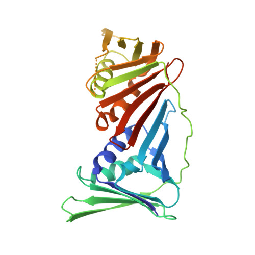
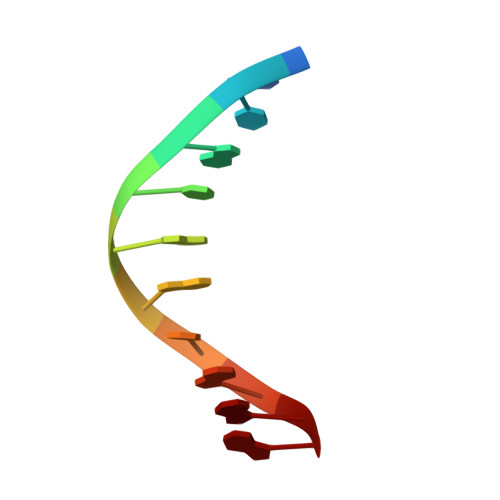
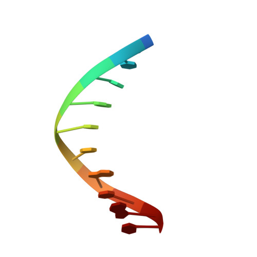
Structural basis of human PCNA sliding on DNA.

De March, M.Merino, N.Barrera-Vilarmau, S.Crehuet, R.Onesti, S.Blanco, F.J.De Biasio, A.

(2017) Nat Commun 8: 13935-13935

  • DOI: https://doi.org/10.1038/ncomms13935
  • Primary Citation of Related Structures:  
    6GIS

  • PubMed Abstract: 

    Sliding clamps encircle DNA and tether polymerases and other factors to the genomic template. However, the molecular mechanism of clamp sliding on DNA is unknown. Using crystallography, NMR and molecular dynamics simulations, here we show that the human clamp PCNA recognizes DNA through a double patch of basic residues within the ring channel, arranged in a right-hand spiral that matches the pitch of B-DNA. We propose that PCNA slides by tracking the DNA backbone via a 'cogwheel' mechanism based on short-lived polar interactions, which keep the orientation of the clamp invariant relative to DNA. Mutation of residues at the PCNA-DNA interface has been shown to impair the initiation of DNA synthesis by polymerase δ (pol δ). Therefore, our findings suggest that a clamp correctly oriented on DNA is necessary for the assembly of a replication-competent PCNA-pol δ holoenzyme.


  • Organizational Affiliation

    Structural Biology Laboratory, Elettra-Sincrotrone Trieste S.C.p.A., 34149 Trieste, Italy.


Macromolecules

Find similar proteins by:  (by identity cutoff)  |  3D Structure
Entity ID: 1
MoleculeChains Sequence LengthOrganismDetailsImage
Proliferating cell nuclear antigen
A, B, C
262Homo sapiensMutation(s): 0 
Gene Names: PCNA
UniProt & NIH Common Fund Data Resources
Find proteins for P12004 (Homo sapiens)
Explore P12004 
Go to UniProtKB:  P12004
PHAROS:  P12004
GTEx:  ENSG00000132646 
Entity Groups  
Sequence Clusters30% Identity50% Identity70% Identity90% Identity95% Identity100% Identity
UniProt GroupP12004
Sequence Annotations
Expand
  • Reference Sequence

Find similar nucleic acids by:  Sequence   |   3D Structure  

Entity ID: 2
MoleculeChains LengthOrganismImage
DNA (5'-D(P*AP*TP*AP*CP*GP*AP*TP*GP*GP*G)-3')10synthetic construct
Sequence Annotations
Expand
  • Reference Sequence

Find similar nucleic acids by:  Sequence   |   3D Structure  

Entity ID: 3
MoleculeChains LengthOrganismImage
DNA (5'-D(P*CP*CP*CP*AP*TP*CP*GP*TP*AP*T)-3')10synthetic construct
Sequence Annotations
Expand
  • Reference Sequence
Experimental Data & Validation

Experimental Data

  • Method: X-RAY DIFFRACTION
  • Resolution: 2.82 Å
  • R-Value Free: 0.284 
  • R-Value Work: 0.247 
  • R-Value Observed: 0.248 
  • Space Group: H 3
Unit Cell:
Length ( Å )Angle ( ˚ )
a = 180.189α = 90
b = 180.189β = 90
c = 76.832γ = 120
Software Package:
Software NamePurpose
REFMACrefinement
XDSdata reduction
SCALAdata scaling
MOLREPphasing

Structure Validation

View Full Validation Report


Entry History 

Deposition Data

Revision History  (Full details and data files)

  • Version 1.0: 2018-05-30
    Type: Initial release
  • Version 2.0: 2019-10-02
    Changes: Advisory, Atomic model, Data collection, Database references, Derived calculations, Other, Polymer sequence, Source and taxonomy, Structure summary
  • Version 2.1: 2024-01-17
    Changes: Data collection, Database references, Refinement description