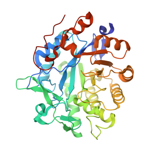
Nonequivalence of Second Sphere "Noncatalytic" Residues in Pentaerythritol Tetranitrate Reductase in Relation to Local Dynamics Linked to H-Transfer in Reactions with NADH and NADPH Coenzymes.
Iorgu, A.I., Baxter, N.J., Cliff, M.J., Levy, C., Waltho, J.P., Hay, S., Scrutton, N.S.(2018) ACS Catal 8: 11589-11599
- PubMed: 31119061 Search on PubMedSearch on PubMed Central
- DOI: https://doi.org/10.1021/acscatal.8b02810
- Primary Citation Related Structures:
6GI7, 6GI8, 6GI9, 6GIA - PubMed Abstract:
Many enzymes that catalyze hydride transfer reactions work via a mechanism dominated by quantum mechanical tunneling. The involvement of fast vibrational modes of the reactive complex is often inferred in these reactions, as in the case of the NAD(P)H-dependent pentaerythritol tetranitrate reductase (PETNR). Herein, we interrogated the H-transfer mechanism in PETNR by designing conservative (L25I and I107L) and side chain shortening (L25A and I107A) PETNR variants and using a combination of experimental approaches (stopped-flow rapid kinetics, X-ray crystallography, isotope/temperature dependence studies of H-transfer and NMR spectroscopy). X-ray data show subtle changes in the local environment of the targeted side chains but no major structural perturbation caused by mutagenesis of these two second sphere active site residues. However, temperature dependence studies of H-transfer revealed a coenzyme-specific and complex thermodynamic equilibrium between different reactive configurations in PETNR-coenzyme complexes. We find that mutagenesis of these second sphere "noncatalytic" residues affects differently the reactivity of PETNR with NADPH and NADH coenzymes. We attribute this to subtle, dynamic structural changes in the PETNR active site, the effects of which impact differently in the nonequivalent reactive geometries of PETNR-NADH and PETNR-NADPH complexes. This inference is confirmed through changes observed in the NMR chemical shift data for PETNR complexes with unreactive 1,4,5,6-tetrahydro-NAD(P) analogues. We show that H-transfer rates can (to some extent) be buffered through entropy-enthalpy compensation, but that use of integrated experimental tools reveals hidden complexities that implicate a role for dynamics in this relatively simple H-transfer reaction. Similar approaches are likely to be informative in other enzymes to understand the relative importance of (distal) hydrophobic side chains and dynamics in controlling the rates of enzymatic H-transfer.
- Manchester Institute of Biotechnology and School of Chemistry, Faculty of Science and Engineering, The University of Manchester, 131 Princess Street, Manchester M1 7DN, United Kingdom.
Organizational Affiliation: