6GGV

Structure of the arginine-bound form of truncated (residues 20-233) ArgBP from T. maritima


Experimental Data Snapshot

  • Method: X-RAY DIFFRACTION
  • Resolution: 2.69 Å
  • R-Value Free: 0.226 
  • R-Value Work: 0.175 
  • R-Value Observed: 0.177 

wwPDB Validation   3D Report Full Report


Ligand Structure Quality Assessment 


This is version 1.2 of the entry. See complete history


Literature

Domain swapping dissection in Thermotoga maritima arginine binding protein: How structural flexibility may compensate destabilization.

Smaldone, G.Berisio, R.Balasco, N.D'Auria, S.Vitagliano, L.Ruggiero, A.

(2018) Biochim Biophys Acta 1866: 952-962

  • DOI: https://doi.org/10.1016/j.bbapap.2018.05.016
  • Primary Citation of Related Structures:  
    6GGP, 6GGV

  • PubMed Abstract: 

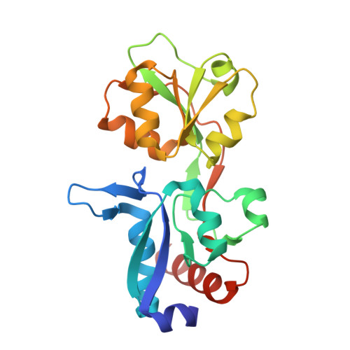
    Thermotoga maritima Arginine Binding Protein (TmArgBP) is a valuable candidate for arginine biosensing in diagnostics. This protein is endowed with unusual structural properties that include an extraordinary thermal/chemical stability, a domain swapped structure that undergoes large tertiary and quaternary structural transition, and the ability to form non-canonical oligomeric species. As the intrinsic stability of TmArgBP allows for extensive protein manipulations, we here dissected its structure in two parts: its main body deprived of the swapping fragment (TmArgBP 20-233 ) and the C-terminal peptide corresponding to the helical swapping element. Both elements have been characterized independently or in combination using a repertoire of biophysical/structural techniques. Present investigations clearly indicate that TmArgBP 20-233 represents a better scaffold for arginine sensing compared to the wild-type protein. Moreover, our data demonstrate that the ligand-free and the ligand-bound forms respond very differently to this helix deletion. This drastic perturbation has an important impact on the ligand-bound form of TmArgBP 20-233 stability whereas it barely affects its ligand-free state. The crystallographic structures of these forms provide a rationale to this puzzling observation. Indeed, the arginine-bound state is very rigid and virtually unchanged upon protein truncation. On the other hand, the flexible ligand-free TmArgBP 20-233 is able to adopt a novel state as a consequence of the helix deletion. Therefore, the flexibility of the ligand-free form endows this state with a remarkable robustness upon severe perturbations. In this scenario, TmArgBP dissection highlights an intriguing connection between destabilizing/stabilizing effects and the overall flexibility that could operate also in other proteins.


  • Organizational Affiliation

    IRCCS SDN, 80143 Napoli, Italy.


Macromolecules
Find similar proteins by:  (by identity cutoff)  |  3D Structure
Entity ID: 1
MoleculeChains Sequence LengthOrganismDetailsImage
Amino acid ABC transporter, periplasmic amino acid-binding protein
A, B
213Thermotoga maritima MSB8Mutation(s): 0 
Gene Names: TM_0593
UniProt
Find proteins for Q9WZ62 (Thermotoga maritima (strain ATCC 43589 / DSM 3109 / JCM 10099 / NBRC 100826 / MSB8))
Explore Q9WZ62 
Go to UniProtKB:  Q9WZ62
Entity Groups  
Sequence Clusters30% Identity50% Identity70% Identity90% Identity95% Identity100% Identity
UniProt GroupQ9WZ62
Sequence Annotations
Expand
  • Reference Sequence
Small Molecules
Ligands 2 Unique
IDChains Name / Formula / InChI Key2D Diagram3D Interactions
ARG
Query on ARG

Download Ideal Coordinates CCD File 
C [auth A],
Q [auth B]
ARGININE
C6 H15 N4 O2
ODKSFYDXXFIFQN-BYPYZUCNSA-O
CD
Query on CD

Download Ideal Coordinates CCD File 
D [auth A]
E [auth A]
F [auth A]
G [auth A]
H [auth A]
D [auth A],
E [auth A],
F [auth A],
G [auth A],
H [auth A],
I [auth A],
J [auth A],
K [auth A],
L [auth A],
M [auth A],
N [auth A],
O [auth A],
P [auth A],
R [auth B],
S [auth B],
T [auth B],
U [auth B],
V [auth B],
W [auth B],
X [auth B],
Y [auth B],
Z [auth B]
CADMIUM ION
Cd
WLZRMCYVCSSEQC-UHFFFAOYSA-N
Experimental Data & Validation

Experimental Data

  • Method: X-RAY DIFFRACTION
  • Resolution: 2.69 Å
  • R-Value Free: 0.226 
  • R-Value Work: 0.175 
  • R-Value Observed: 0.177 
  • Space Group: P 21 2 21
Unit Cell:
Length ( Å )Angle ( ˚ )
a = 30.7α = 90
b = 52.139β = 90
c = 221.343γ = 90
Software Package:
Software NamePurpose
REFMACrefinement
HKL-2000data reduction
HKL-2000data scaling
PHASERphasing

Structure Validation

View Full Validation Report


Ligand Structure Quality Assessment 


Entry History 

Deposition Data

Revision History  (Full details and data files)

  • Version 1.0: 2018-06-13
    Type: Initial release
  • Version 1.1: 2018-06-20
    Changes: Data collection, Database references
  • Version 1.2: 2024-01-17
    Changes: Data collection, Database references, Derived calculations, Refinement description