Molecular basis for AU-rich element recognition and dimerization by the HuR C-terminal RRM.
Ripin, N., Boudet, J., Duszczyk, M.M., Hinniger, A., Faller, M., Krepl, M., Gadi, A., Schneider, R.J., Sponer, J., Meisner-Kober, N.C., Allain, F.H.(2019) Proc Natl Acad Sci U S A 116: 2935-2944
- PubMed: 30718402 Search on PubMedSearch on PubMed Central
- DOI: https://doi.org/10.1073/pnas.1808696116
- Primary Citation Related Structures:
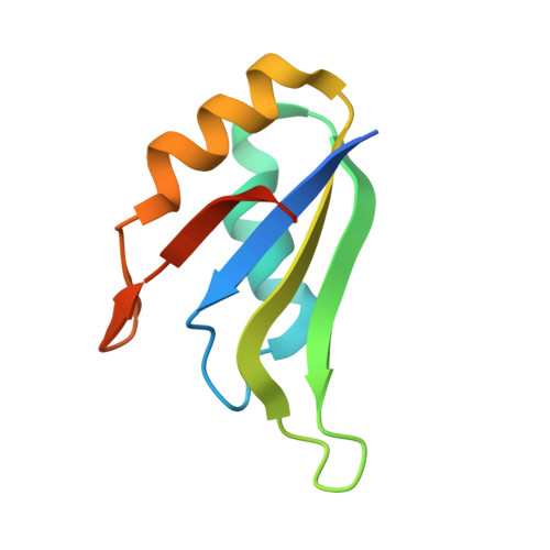
6GC5 - PubMed Abstract:
Human antigen R (HuR) is a key regulator of cellular mRNAs containing adenylate/uridylate-rich elements (AU-rich elements; AREs). These are a major class of cis elements within 3' untranslated regions, targeting these mRNAs for rapid degradation. HuR contains three RNA recognition motifs (RRMs): a tandem RRM1 and 2, followed by a flexible linker and a C-terminal RRM3. While RRM1 and 2 are structurally characterized, little is known about RRM3. Here we present a 1.9-Å-resolution crystal structure of RRM3 bound to different ARE motifs. This structure together with biophysical methods and cell-culture assays revealed the mechanism of RRM3 ARE recognition and dimerization. While multiple RNA motifs can be bound, recognition of the canonical AUUUA pentameric motif is possible by binding to two registers. Additionally, RRM3 forms homodimers to increase its RNA binding affinity. Finally, although HuR stabilizes ARE-containing RNAs, we found that RRM3 counteracts this effect, as shown in a cell-based ARE reporter assay and by qPCR with native HuR mRNA targets containing multiple AUUUA motifs, possibly by competing with RRM12.
- Department of Biology, Institute of Molecular Biology and Biophysics, ETH Zürich, 8093 Zürich, Switzerland; nina.ripin@mol.biol.ethz.ch nicole.meisner-kober@sbg.ac.at frederic.allain@mol.biol.ethz.ch.
Organizational Affiliation: