6G4G | pdb_00006g4g

Full length ectodomain of ectonucleotide phosphodiesterase/pyrophosphatase-3 (NPP3) including the SMB domains but with a partially disordered active site structure


Experimental Data Snapshot

  • Method: X-RAY DIFFRACTION
  • Resolution: 2.80 Å
  • R-Value Free: 
    0.237 (Depositor), 0.251 (DCC) 
  • R-Value Work: 
    0.196 (Depositor), 0.209 (DCC) 
  • R-Value Observed: 
    0.198 (Depositor) 

Starting Model: experimental
View more details

wwPDB Validation 3D Report Full Report

Validation slider image for 6G4G

Ligand Structure Quality Assessment 


This is version 2.2 of the entry. See complete history

Literature

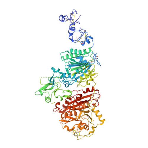
Crystallization of ectonucleotide phosphodiesterase/pyrophosphatase-3 and orientation of the SMB domains in the full-length ectodomain.

Dohler, C.Zebisch, M.Krinke, D.Robitzki, A.Strater, N.

(2018) Acta Crystallogr F Struct Biol Commun 74: 696-703

  • DOI: https://doi.org/10.1107/S2053230X18011111
  • Primary Citation Related Structures: 
    6G4G

  • PubMed Abstract: 

    Ectonucleotide phosphodiesterase/pyrophosphatase-3 (NPP3, ENPP3) is an ATP-hydrolyzing glycoprotein that is located in the extracellular space. The full-length ectodomain of rat NPP3 was expressed in HEK293S GntI - cells, purified using two chromatographic steps and crystallized. Its structure at 2.77 Å resolution reveals that the active-site zinc ions are missing and a large part of the active site and the surrounding residues are flexible. The SMB-like domains have the same orientation in all four molecules in the asymmetric unit. The SMB2 domain is oriented as in NPP2, but the SMB1 domain does not interact with the PDE domain but extends further away from the PDE domain. Deletion of the SMB domains resulted in crystals that diffracted to 2.4 Å resolution and are suitable for substrate-binding studies.


  • Organizational Affiliation
    • Institute of Bioanalytical Chemistry, Center for Biotechnology and Biomedicine, Leipzig University, Deutscher Platz 5, 04103 Leipzig, Germany.

Macromolecule Content 

  • Total Structure Weight: 387.2 kDa 
  • Atom Count: 24,786 
  • Modeled Residue Count: 3,047 
  • Deposited Residue Count: 3,360 
  • Unique protein chains: 1

Macromolecules

Find similar proteins by:|  3D Structure
Entity ID: 1
MoleculeChains  Sequence LengthOrganismDetailsImage
Ectonucleotide pyrophosphatase/phosphodiesterase family member 3
A, B, C, D
840Rattus norvegicusMutation(s): 3 
Gene Names: Enpp3Pdnp3
EC: 3.1.4.1 (PDB Primary Data), 3.6.1.9 (PDB Primary Data), 3.6.1 (UniProt)
UniProt
Find proteins for P97675 (Rattus norvegicus)
Explore P97675 
Go to UniProtKB:  P97675
Entity Groups
Sequence Clusters30% Identity50% Identity70% Identity90% Identity95% Identity100% Identity
UniProt GroupP97675
Glycosylation
Glycosylation Sites: 4Go to GlyGen: P97675-1
Sequence Annotations
Expand
Reference Sequence

Oligosaccharides

Help  
Entity ID: 2
MoleculeChains Length2D Diagram GlycosylationD Interactions
2-acetamido-2-deoxy-beta-D-glucopyranose-(1-4)-2-acetamido-2-deoxy-beta-D-glucopyranose
E, F, G, H, I
E, F, G, H, I, J, K
2N-Glycosylation
Glycosylation Resources
GlyTouCan: G42666HT
GlyCosmos: G42666HT
GlyGen: G42666HT

Small Molecules

Ligands 2 Unique
IDChains Name / Formula / InChI Key2D Diagram3D Interactions
NAG

Query on NAG


Download:Ideal Coordinates CCD File
M [auth A]
N [auth A]
O [auth A]
Q [auth B]
R [auth B]
M [auth A],
N [auth A],
O [auth A],
Q [auth B],
R [auth B],
T [auth C],
V [auth D],
W [auth D]
2-acetamido-2-deoxy-beta-D-glucopyranose
C8 H15 N O6
OVRNDRQMDRJTHS-FMDGEEDCSA-N
CA

Query on CA


Download:Ideal Coordinates CCD File
L [auth A],
P [auth B],
S [auth C],
U [auth D]
CALCIUM ION
Ca
BHPQYMZQTOCNFJ-UHFFFAOYSA-N

Experimental Data & Validation

Experimental Data

  • Method: X-RAY DIFFRACTION
  • Resolution: 2.80 Å
  • R-Value Free:  0.237 (Depositor), 0.251 (DCC) 
  • R-Value Work:  0.196 (Depositor), 0.209 (DCC) 
  • R-Value Observed: 0.198 (Depositor) 
Space Group: P 1
Unit Cell:
Length ( Å )Angle ( ˚ )
a = 76.36α = 86.77
b = 116.3β = 87.76
c = 124.16γ = 88.2
Software Package:
Software NamePurpose
BUSTERrefinement
DIALSdata reduction
STARANISOdata scaling
Aimlessdata scaling
PHASERphasing

Structure Validation

View Full Validation Report


Ligand Structure Quality Assessment 


Entry History 

& Funding Information

Deposition Data


Funding OrganizationLocationGrant Number
German Research FoundationGermany477/13-2

Revision History  (Full details and data files)

  • Version 1.0: 2018-11-07
    Type: Initial release
  • Version 1.1: 2018-11-14
    Changes: Data collection, Database references
  • Version 2.0: 2020-07-29
    Type: Remediation
    Reason: Carbohydrate remediation
    Changes: Atomic model, Data collection, Derived calculations, Structure summary
  • Version 2.1: 2024-01-17
    Changes: Data collection, Database references, Refinement description, Structure summary
  • Version 2.2: 2024-10-23
    Changes: Structure summary