6G47

Crystal Structure of Human Adenovirus 52 Short Fiber Knob in Complex with alpha-(2,8)-Trisialic Acid (DP3)


Experimental Data Snapshot

  • Method: X-RAY DIFFRACTION
  • Resolution: 1.50 Å
  • R-Value Free: 0.178 
  • R-Value Work: 0.162 
  • R-Value Observed: 0.163 

wwPDB Validation   3D Report Full Report


Ligand Structure Quality Assessment 


This is version 2.1 of the entry. See complete history


Literature

Polysialic acid is a cellular receptor for human adenovirus 52.

Lenman, A.Liaci, A.M.Liu, Y.Frangsmyr, L.Frank, M.Blaum, B.S.Chai, W.Podgorski, I.I.Harrach, B.Benko, M.Feizi, T.Stehle, T.Arnberg, N.

(2018) Proc Natl Acad Sci U S A 115: E4264-E4273

  • DOI: https://doi.org/10.1073/pnas.1716900115
  • Primary Citation of Related Structures:  
    6G47

  • PubMed Abstract: 

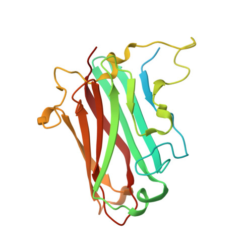
    Human adenovirus 52 (HAdV-52) is one of only three known HAdVs equipped with both a long and a short fiber protein. While the long fiber binds to the coxsackie and adenovirus receptor, the function of the short fiber in the virus life cycle is poorly understood. Here, we show, by glycan microarray analysis and cellular studies, that the short fiber knob (SFK) of HAdV-52 recognizes long chains of α-2,8-linked polysialic acid (polySia), a large posttranslational modification of selected carrier proteins, and that HAdV-52 can use polySia as a receptor on target cells. X-ray crystallography, NMR, molecular dynamics simulation, and structure-guided mutagenesis of the SFK reveal that the nonreducing, terminal sialic acid of polySia engages the protein with direct contacts, and that specificity for polySia is achieved through subtle, transient electrostatic interactions with additional sialic acid residues. In this study, we present a previously unrecognized role for polySia as a cellular receptor for a human viral pathogen. Our detailed analysis of the determinants of specificity for this interaction has general implications for protein-carbohydrate interactions, particularly concerning highly charged glycan structures, and provides interesting dimensions on the biology and evolution of members of Human mastadenovirus G .


  • Organizational Affiliation

    Division of Virology, Department of Clinical Microbiology, and Laboratory for Molecular Infection Medicine Sweden, Umeå University, SE-90185 Umeå, Sweden; annasara.lenman@umu.se thilo.stehle@uni-tuebingen.de.


Macromolecules
Find similar proteins by:  (by identity cutoff)  |  3D Structure
Entity ID: 1
MoleculeChains Sequence LengthOrganismDetailsImage
Fiber-1
A, B, C
209human adenovirus 52Mutation(s): 0 
UniProt
Find proteins for A0MK70 (Human adenovirus 52)
Explore A0MK70 
Go to UniProtKB:  A0MK70
Entity Groups  
Sequence Clusters30% Identity50% Identity70% Identity90% Identity95% Identity100% Identity
UniProt GroupA0MK70
Sequence Annotations
Expand
  • Reference Sequence
Oligosaccharides

Help

Entity ID: 2
MoleculeChains Length2D Diagram Glycosylation3D Interactions
N-acetyl-alpha-neuraminic acid-(2-8)-N-acetyl-alpha-neuraminic acid
D
2N/A
Glycosylation Resources
GlyTouCan:  G87492XO
GlyCosmos:  G87492XO
GlyGen:  G87492XO
Small Molecules
Ligands 4 Unique
IDChains Name / Formula / InChI Key2D Diagram3D Interactions
SIA (Subject of Investigation/LOI)
Query on SIA

Download Ideal Coordinates CCD File 
E [auth A]N-acetyl-alpha-neuraminic acid
C11 H19 N O9
SQVRNKJHWKZAKO-YRMXFSIDSA-N
TRS
Query on TRS

Download Ideal Coordinates CCD File 
I [auth C]2-AMINO-2-HYDROXYMETHYL-PROPANE-1,3-DIOL
C4 H12 N O3
LENZDBCJOHFCAS-UHFFFAOYSA-O
MRD
Query on MRD

Download Ideal Coordinates CCD File 
G [auth B],
H [auth B]
(4R)-2-METHYLPENTANE-2,4-DIOL
C6 H14 O2
SVTBMSDMJJWYQN-RXMQYKEDSA-N
MPD
Query on MPD

Download Ideal Coordinates CCD File 
F [auth A],
J [auth C]
(4S)-2-METHYL-2,4-PENTANEDIOL
C6 H14 O2
SVTBMSDMJJWYQN-YFKPBYRVSA-N
Experimental Data & Validation

Experimental Data

  • Method: X-RAY DIFFRACTION
  • Resolution: 1.50 Å
  • R-Value Free: 0.178 
  • R-Value Work: 0.162 
  • R-Value Observed: 0.163 
  • Space Group: P 21 21 21
Unit Cell:
Length ( Å )Angle ( ˚ )
a = 64.88α = 90
b = 81.71β = 90
c = 93.53γ = 90
Software Package:
Software NamePurpose
PHENIXrefinement
PDB_EXTRACTdata extraction
XDSdata reduction
XSCALEdata scaling
MOLREPphasing

Structure Validation

View Full Validation Report


Ligand Structure Quality Assessment 


Entry History & Funding Information

Deposition Data


Funding OrganizationLocationGrant Number
German Research FoundationGermanySFB 685

Revision History  (Full details and data files)

  • Version 1.0: 2018-05-02
    Type: Initial release
  • Version 1.1: 2018-05-09
    Changes: Data collection, Database references
  • Version 2.0: 2020-07-29
    Type: Remediation
    Reason: Carbohydrate remediation
    Changes: Atomic model, Data collection, Derived calculations, Structure summary
  • Version 2.1: 2024-01-17
    Changes: Data collection, Database references, Derived calculations, Refinement description, Structure summary