6G0W

Human PARP14 (ARTD8), catalytic fragment in complex with inhibitor MCD72


Experimental Data Snapshot

  • Method: X-RAY DIFFRACTION
  • Resolution: 2.34 Å
  • R-Value Free: 0.275 
  • R-Value Work: 0.224 
  • R-Value Observed: 0.226 

wwPDB Validation   3D Report Full Report


Ligand Structure Quality Assessment 


This is version 1.2 of the entry. See complete history


Literature

Design, synthesis and evaluation of potent and selective inhibitors of mono-(ADP-ribosyl)transferases PARP10 and PARP14.

Holechek, J.Lease, R.Thorsell, A.G.Karlberg, T.McCadden, C.Grant, R.Keen, A.Callahan, E.Schuler, H.Ferraris, D.

(2018) Bioorg Med Chem Lett 28: 2050-2054

  • DOI: https://doi.org/10.1016/j.bmcl.2018.04.056
  • Primary Citation of Related Structures:  
    6G0W

  • PubMed Abstract: 

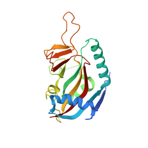
    A series of diaryl ethers were designed and synthesized to discern the structure activity relationships against the two closely related mono-(ADP-ribosyl)transferases PARP10 and PARP14. Structure activity studies identified 8b as a sub-micromolar inhibitor of PARP10 with ∼15-fold selectivity over PARP14. In addition, 8k and 8m were discovered to have sub-micromolar potency against PARP14 and demonstrated moderate selectivity over PARP10. A crystal structure of the complex of PARP14 and 8b shows binding of the compound in a novel hydrophobic pocket and explains both potency and selectivity over other PARP family members. In addition, 8b, 8k and 8m also demonstrate selectivity over PARP1. Together, this study identified novel, potent and metabolically stable derivatives to use as chemical probes for these biologically interesting therapeutic targets.


  • Organizational Affiliation

    McDaniel College, 2 College Hill, Westminster, MD 21157, United States.


Macromolecules
Find similar proteins by:  (by identity cutoff)  |  3D Structure
Entity ID: 1
MoleculeChains Sequence LengthOrganismDetailsImage
Poly [ADP-ribose] polymerase 14
A, B
193Homo sapiensMutation(s): 0 
Gene Names: PARP14BAL2KIAA1268
EC: 2.4.2.30
UniProt & NIH Common Fund Data Resources
Find proteins for Q460N5 (Homo sapiens)
Explore Q460N5 
Go to UniProtKB:  Q460N5
PHAROS:  Q460N5
GTEx:  ENSG00000173193 
Entity Groups  
Sequence Clusters30% Identity50% Identity70% Identity90% Identity95% Identity100% Identity
UniProt GroupQ460N5
Sequence Annotations
Expand
  • Reference Sequence
Small Molecules
Ligands 1 Unique
IDChains Name / Formula / InChI Key2D Diagram3D Interactions
EGW
Query on EGW

Download Ideal Coordinates CCD File 
C [auth A],
D [auth B]
4-[3-[4-(4-fluorophenyl)piperidin-1-yl]carbonylphenoxy]benzamide
C25 H23 F N2 O3
GKQVMMKXPPAKMY-UHFFFAOYSA-N
Binding Affinity Annotations 
IDSourceBinding Affinity
EGW Binding MOAD:  6G0W IC50: 5200 (nM) from 1 assay(s)
BindingDB:  6G0W IC50: min: 5200, max: 5248 (nM) from 2 assay(s)
Experimental Data & Validation

Experimental Data

  • Method: X-RAY DIFFRACTION
  • Resolution: 2.34 Å
  • R-Value Free: 0.275 
  • R-Value Work: 0.224 
  • R-Value Observed: 0.226 
  • Space Group: P 61 2 2
Unit Cell:
Length ( Å )Angle ( ˚ )
a = 83.314α = 90
b = 83.314β = 90
c = 255.495γ = 120
Software Package:
Software NamePurpose
PHENIXrefinement
XDSdata reduction
XSCALEdata scaling
PHASERphasing

Structure Validation

View Full Validation Report


Ligand Structure Quality Assessment 


Entry History 

Deposition Data

Revision History  (Full details and data files)

  • Version 1.0: 2018-05-23
    Type: Initial release
  • Version 1.1: 2018-05-30
    Changes: Data collection, Database references
  • Version 1.2: 2024-01-17
    Changes: Data collection, Database references, Derived calculations, Refinement description