6FZN

SMURFP-Y56R mutant in complex with biliverdin


Experimental Data Snapshot

  • Method: X-RAY DIFFRACTION
  • Resolution: 2.50 Å
  • R-Value Free: 0.263 
  • R-Value Work: 0.212 
  • R-Value Observed: 0.215 

wwPDB Validation   3D Report Full Report


Ligand Structure Quality Assessment 


This is version 1.2 of the entry. See complete history


Literature

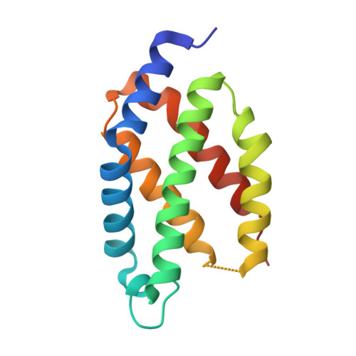
Crystal structure of a biliverdin-bound phycobiliprotein: Interdependence of oligomerization and chromophorylation.

Fuenzalida-Werner, J.P.Janowski, R.Mishra, K.Weidenfeld, I.Niessing, D.Ntziachristos, V.Stiel, A.C.

(2018) J Struct Biol 204: 519-522

  • DOI: https://doi.org/10.1016/j.jsb.2018.09.013
  • Primary Citation of Related Structures:  
    6FZN, 6FZO

  • PubMed Abstract: 

    Small, ultra-red fluorescence protein (smURFP) introduces the non-native biliverdin (BV) chromophore to phycobiliproteins (PBPs), allowing them to be used as transgenic labels for in vivo mammalian imaging. Presently, no structural information exists for PBPs bound to the non-native BV chromophore, which limits the further development of smURFP and related proteins as imaging labels or indicators. Here we describe the first crystal structure of a PBP bound to BV. The structures of smURFP-Y56R with BV and smURFP-Y56F without BV reveal unique oligomerization interfaces different from those in wild-type PBPs bound to native chromophores. Our structures suggest that the oligomerization interface affects the BV binding site, creating a link between oligomerization and chromophorylation that we confirmed through site-directed mutagenesis and that may help guide efforts to improve the notorious chromophorylation of smURFP and other PBPs engineered to bind BV.


  • Organizational Affiliation

    Institute of Biological and Medical Imaging (IBMI), Helmholtz Zentrum München, Neuherberg, Germany.


Macromolecules
Find similar proteins by:  (by identity cutoff)  |  3D Structure
Entity ID: 1
MoleculeChains Sequence LengthOrganismDetailsImage
smURFP
A, B
138synthetic constructMutation(s): 0 
Entity Groups  
Sequence Clusters30% Identity50% Identity70% Identity90% Identity95% Identity100% Identity
Sequence Annotations
Expand
  • Reference Sequence
Small Molecules
Ligands 1 Unique
IDChains Name / Formula / InChI Key2D Diagram3D Interactions
9UY
Query on 9UY

Download Ideal Coordinates CCD File 
C [auth A]3-[5-[[(3~{R},4~{R})-3-ethenyl-4-methyl-5-oxidanylidene-3,4-dihydropyrrol-2-yl]methyl]-2-[[5-[(4-ethenyl-3-methyl-5-oxidanylidene-pyrrol-2-yl)methyl]-3-(3-hydroxy-3-oxopropyl)-4-methyl-1~{H}-pyrrol-2-yl]methyl]-4-methyl-1~{H}-pyrrol-3-yl]propanoic acid
C33 H38 N4 O6
IAVYOZQSRLDKLE-WOJBJXKFSA-N
Experimental Data & Validation

Experimental Data

  • Method: X-RAY DIFFRACTION
  • Resolution: 2.50 Å
  • R-Value Free: 0.263 
  • R-Value Work: 0.212 
  • R-Value Observed: 0.215 
  • Space Group: P 1 21 1
Unit Cell:
Length ( Å )Angle ( ˚ )
a = 42.55α = 90
b = 57.96β = 105.04
c = 52.11γ = 90
Software Package:
Software NamePurpose
REFMACrefinement
XDSdata reduction
XSCALEdata scaling
Auto-Rickshawphasing

Structure Validation

View Full Validation Report


Ligand Structure Quality Assessment 


Entry History 

Revision History  (Full details and data files)

  • Version 1.0: 2018-10-17
    Type: Initial release
  • Version 1.1: 2018-12-26
    Changes: Data collection, Database references
  • Version 1.2: 2019-02-20
    Changes: Data collection, Source and taxonomy