6FYJ

Cytochrome P450 peroxygenase CYP152K6 in complex with Myristic Acid


Experimental Data Snapshot

  • Method: X-RAY DIFFRACTION
  • Resolution: 1.30 Å
  • R-Value Free: 0.193 
  • R-Value Work: 0.173 
  • R-Value Observed: 0.174 

wwPDB Validation   3D Report Full Report


Ligand Structure Quality Assessment 


This is version 1.1 of the entry. See complete history


Literature

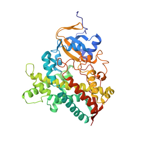
Structural and catalytic properties of the peroxygenase P450 enzyme CYP152K6 from Bacillus methanolicus.

Girvan, H.M.Poddar, H.McLean, K.J.Nelson, D.R.Hollywood, K.A.Levy, C.W.Leys, D.Munro, A.W.

(2018) J Inorg Biochem 188: 18-28

  • DOI: 10.1016/j.jinorgbio.2018.08.002
  • Primary Citation of Related Structures:  
    6FYJ

  • PubMed Abstract: 
  • The CYP152 family of cytochrome P450 enzymes (P450s or CYPs) are bacterial peroxygenases that use hydrogen peroxide to drive hydroxylation and decarboxylation of fatty acid substrates. We have expressed and purified a novel CYP152 family member - CYP152K6 from the methylotroph Bacillus methanolicus MGA3 ...

    The CYP152 family of cytochrome P450 enzymes (P450s or CYPs) are bacterial peroxygenases that use hydrogen peroxide to drive hydroxylation and decarboxylation of fatty acid substrates. We have expressed and purified a novel CYP152 family member - CYP152K6 from the methylotroph Bacillus methanolicus MGA3. CYP152K6 was characterized using spectroscopic, analytical and structural methods. CYP152K6, like its peroxygenase counterpart P450 SPα (CYP152B1) from Sphingomonas paucimobilis, does not undergo significant fatty acid-induced perturbation to the heme spectrum, with the exception of a minor Soret shift observed on binding dodecanoic acid. However, CYP152K6 purified from an E. coli expression system was crystallized and its structure was determined to 1.3 Å with tetradecanoic acid bound. No lipids were present in conditions used for crystallogenesis, and thus CYP152K6 must form a complex by incorporating the fatty acid from E. coli cells. Turnover studies with dodecanoic acid revealed several products, with 2-hydroxydodecanoic acid as the major product and much smaller quantities of 3-hydroxydodecanoic acid. Secondary turnover products were undec-1-en-1-ol, 2-hydroxydodec-2-enoic acid and 2,3-dihydroxydodecanoic acid. This is the first report of a 2,3-hydroxylated fatty acid product made by a peroxygenase P450, with the dihydroxylated product formed by CYP152K6-catalyzed 3-hydroxylation of 2-hydroxydodecanoic acid, but not by 2-hydroxylation of 3-hydroxydodecanoic acid.


    Organizational Affiliation

    Centre for Synthetic Biology of Fine and Specialty Chemicals (SYNBIOCHEM), Manchester Institute of Biotechnology, School of Chemistry, The University of Manchester, Manchester M1 7DN, United Kingdom. Electronic address: Andrew.Munro@Manchester.ac.uk.


Macromolecules
Find similar proteins by:  (by identity cutoff)  |  3D Structure
Entity ID: 1
MoleculeChainsSequence LengthOrganismDetailsImage
Fatty-acid peroxygenaseA421Bacillus methanolicusMutation(s): 0 
Gene Names: cypCBMMGA3_06595
EC: 1.11.2.4
UniProt
Find proteins for I3DZK9 (Bacillus methanolicus (strain MGA3 / ATCC 53907))
Explore I3DZK9 
Go to UniProtKB:  I3DZK9
Entity Groups  
Sequence Clusters30% Identity50% Identity70% Identity90% Identity95% Identity100% Identity
UniProt GroupI3DZK9
Protein Feature View
Expand
  • Reference Sequence
Small Molecules
Ligands 3 Unique
IDChainsName / Formula / InChI Key2D Diagram3D Interactions
HEM (Subject of Investigation/LOI)
Query on HEM

Download Ideal Coordinates CCD File 
C [auth A]PROTOPORPHYRIN IX CONTAINING FE
C34 H32 Fe N4 O4
KABFMIBPWCXCRK-RGGAHWMASA-L
 Ligand Interaction
MYR (Subject of Investigation/LOI)
Query on MYR

Download Ideal Coordinates CCD File 
B [auth A]MYRISTIC ACID
C14 H28 O2
TUNFSRHWOTWDNC-UHFFFAOYSA-N
 Ligand Interaction
PEG
Query on PEG

Download Ideal Coordinates CCD File 
D [auth A]DI(HYDROXYETHYL)ETHER
C4 H10 O3
MTHSVFCYNBDYFN-UHFFFAOYSA-N
 Ligand Interaction
Experimental Data & Validation

Experimental Data

  • Method: X-RAY DIFFRACTION
  • Resolution: 1.30 Å
  • R-Value Free: 0.193 
  • R-Value Work: 0.173 
  • R-Value Observed: 0.174 
  • Space Group: C 1 2 1
Unit Cell:
Length ( Å )Angle ( ˚ )
a = 93.952α = 90
b = 71.173β = 103.58
c = 68.425γ = 90
Software Package:
Software NamePurpose
PHENIXrefinement
xia2data reduction
DIALSdata scaling
PHASERphasing

Structure Validation

View Full Validation Report


Ligand Structure Quality Assessment 


Entry History & Funding Information

Deposition Data


Funding OrganizationLocationGrant Number
Biotechnology and Biological Sciences Research CouncilUnited Kingdom--

Revision History  (Full details and data files)

  • Version 1.0: 2018-08-22
    Type: Initial release
  • Version 1.1: 2018-08-29
    Changes: Data collection, Database references